
Complete18MODELS
Eye
models, complete down to the molecule
Visualisy · Medical 3D Studio · Organ → Molecule
Our models don't stop at the shape of an organ. They reveal tissue, cell, organelle and molecule in one connected 3D atlas. We review the science and cross-check every number against independent sources. From the same models, we create your cinematic images and videos.
© Visualisy owns full rights to every model, image and video
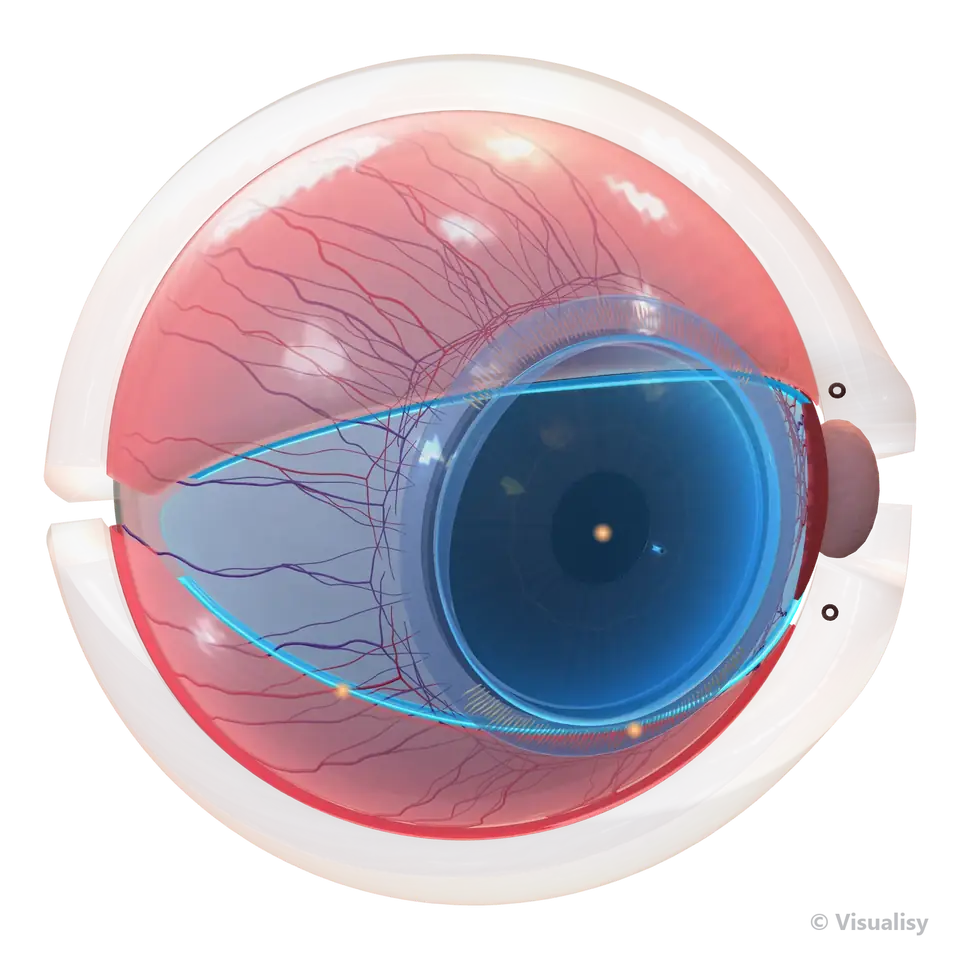
parts in the Eye Atlas
levels: organ, tissue, cell, organelle, molecule
independent sources for every number
scenes in IDRAK 3D
One method · 14 specialties
The Eye Atlas is complete. The rest are planned, with prospective counts that can grow. Models ship with ready-made motion, and advanced motion follows once the science is locked.
BenefitStart with the complete Eye Atlas, or commission the atlas your project needs.
models, complete down to the molecule
models planned, and the count can grow
models planned, and the count can grow
models planned, and the count can grow
models planned, and the count can grow
models planned, and the count can grow
models planned, and the count can grow
models planned, and the count can grow
models planned, and the count can grow
models planned, and the count can grow
models planned, and the count can grow
models planned, and the count can grow
models planned, and the count can grow
models planned, and the count can grow
Start with the ready Eye Atlas, or request an atlas for your specialty, from organ to molecule.
One atlas · products for your project
Our scientific models belong to Visualisy. From them we build your model, your drug and research explainers, your clinic app, your teaching material, and your images and videos, for your website, app, conference or clinic screen.
BenefitCut build time and cost with a ready, reviewed scientific foundation.
Show the drug's journey, how it binds its target and what follows, down to cell and molecule, for conferences, sales teams and physician education.
We build a new structure, a disease state or a medical device on top of the atlas models, using the same scientific method.
Show the mechanism you study or your findings with a scientific model, for a published figure or a conference talk.
Explain the condition with educational models on the clinic screen, and control the view from a tablet.
Discover IDRAK 3D ↓Connect organ to tissue, cell and molecule in one continuous view, at the depth your curriculum needs.
Images and videos from our original models, with full copyright owned by Visualisy and a licence that defines how you use them in your project.
See the production stages ↓And alsoIllustrative visual simulationMedical device demosSurgical step walkthroughsExplaining options before decisionsVirtual and augmented reality
Save build time and cost: we prepare your project deliverables from scientific models owned by Visualisy.
Case study · Eye Atlas
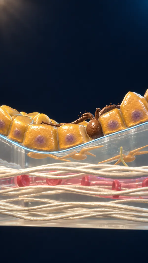
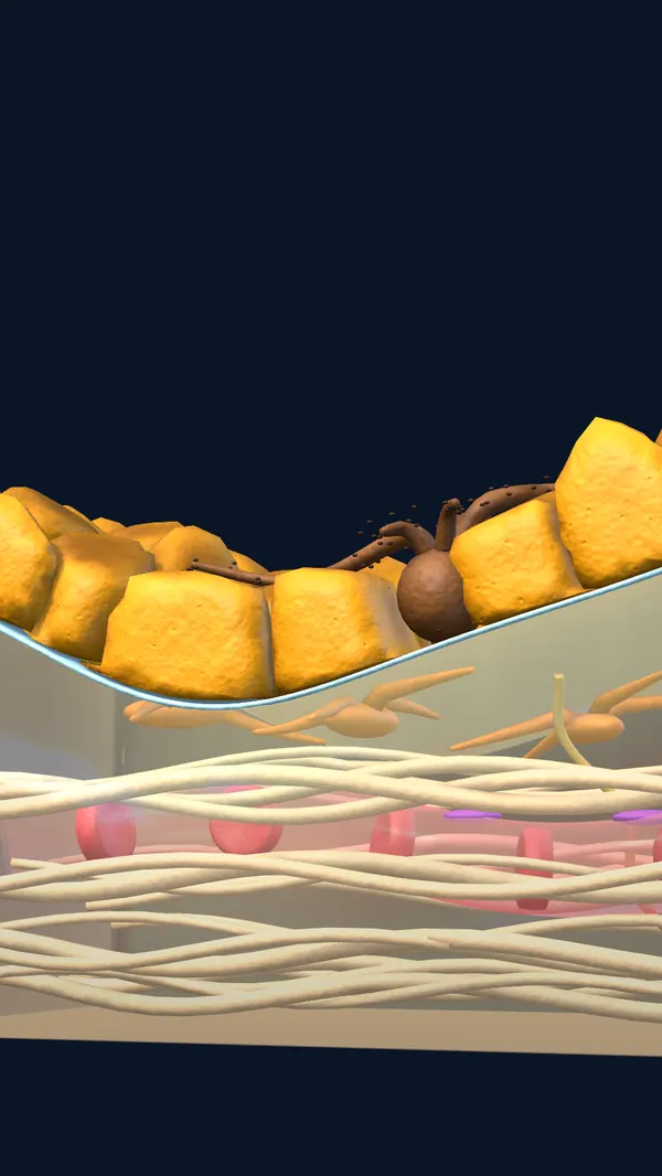
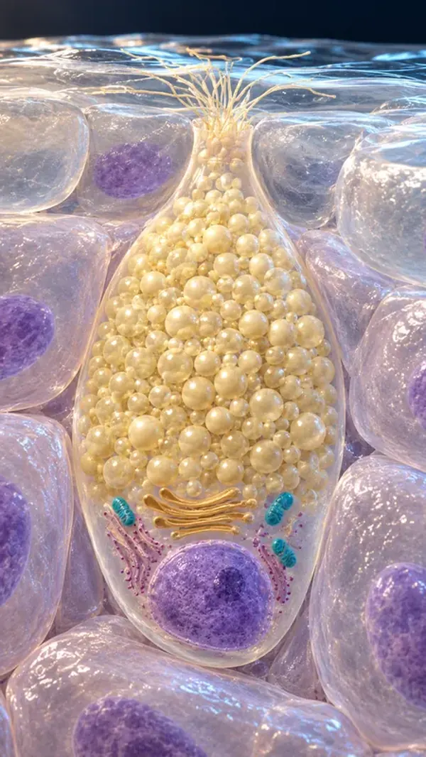
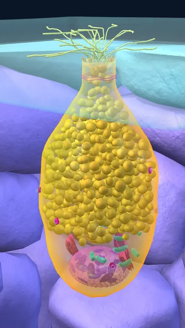
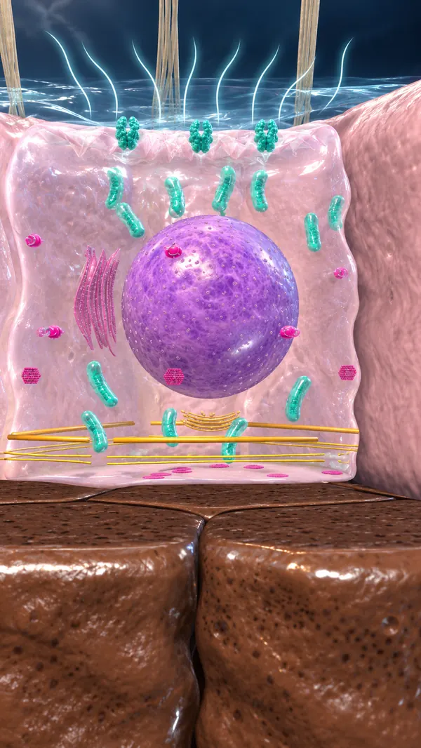
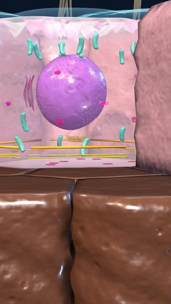
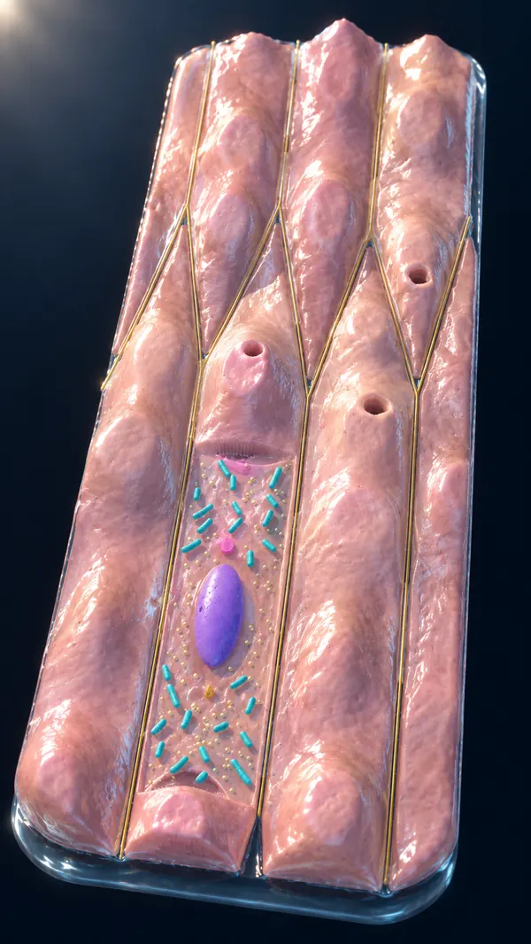
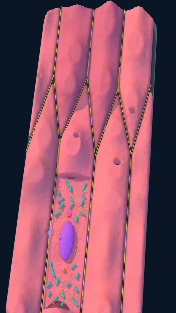
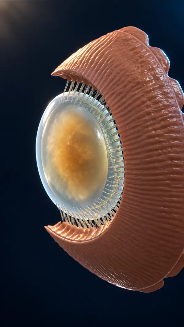
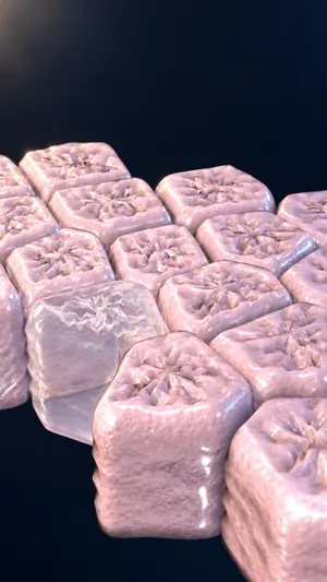
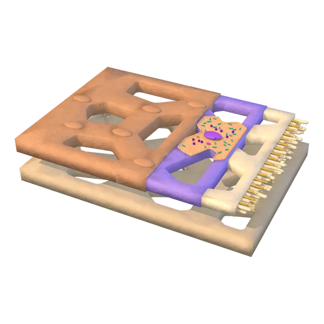
The Eye Atlas has 18 parts, each its own model with magnification levels. Here is one example from the ocular surface.
BenefitLink what you see in the organ to what happens at the cell and molecule level.
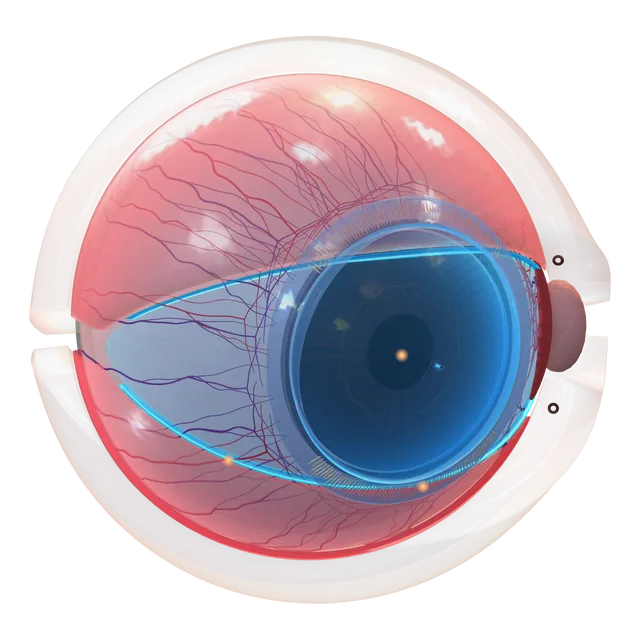
Ocular surface
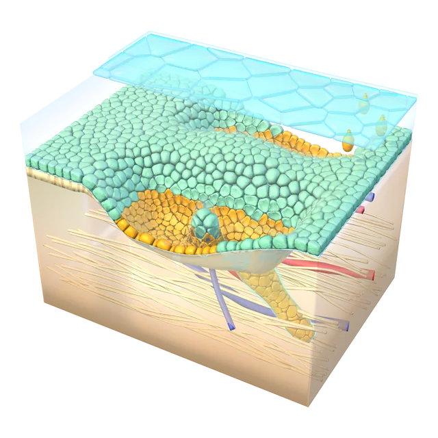
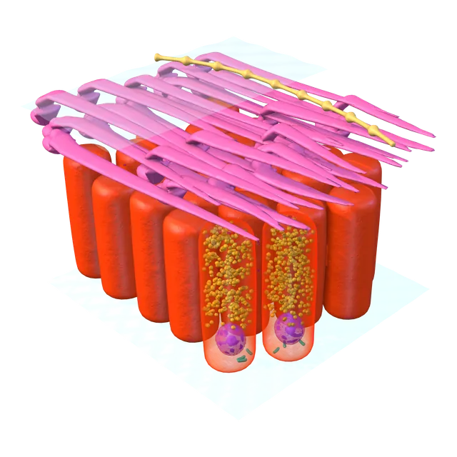
Limbus and stem cell niche
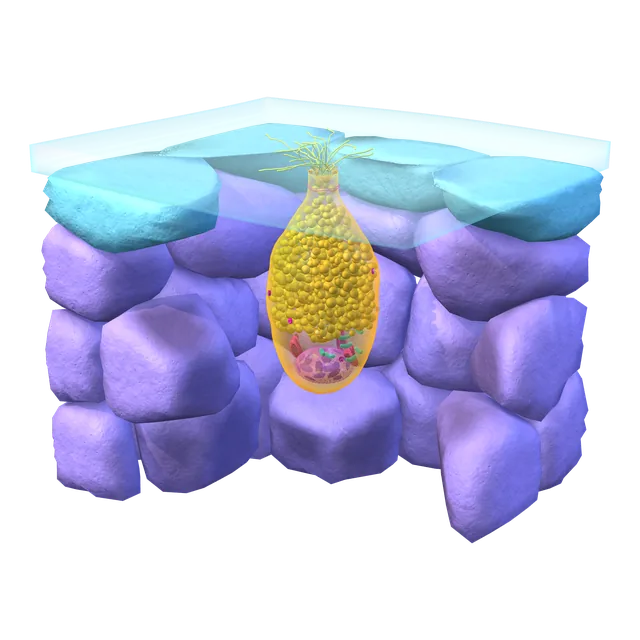
Goblet cell
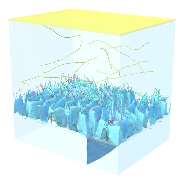
Microplicae and glycocalyx
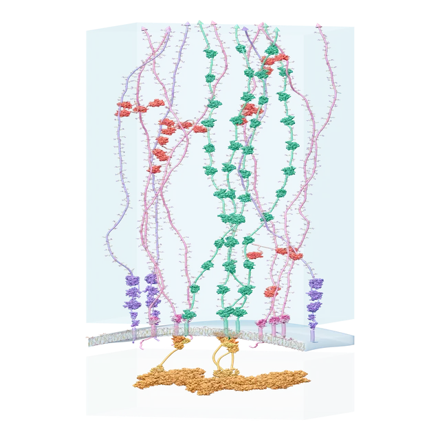
Mucins and glycans on the cell surface
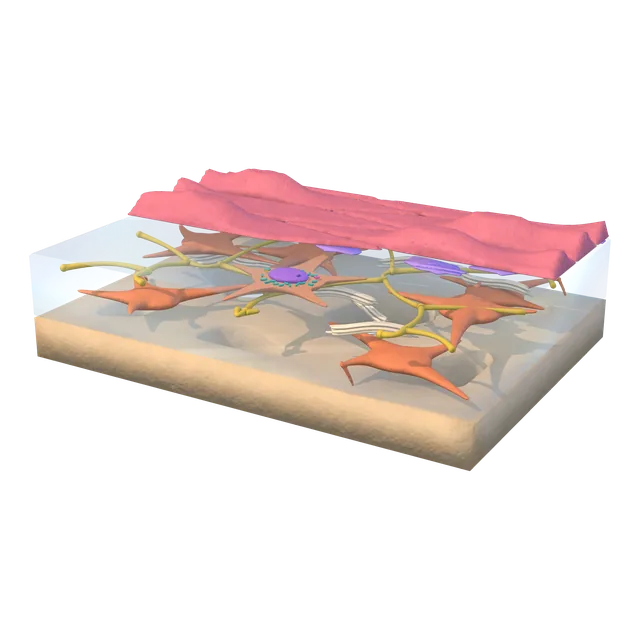
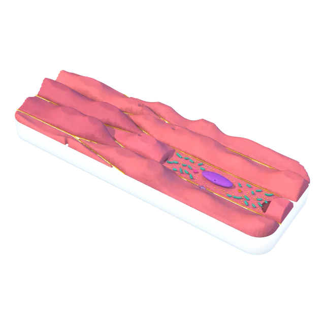
Explore more: the cornea down to collagen fibrils, the eye muscles down to the sarcomere, and the optic nerve down to the node of Ranvier.
Explain what happens inside the organ, down to cell and molecule, at the depth your audience needs.
Atlas Cinematic · Model → image → motion
From our scientific models, we use AI to create a cinematic image with the same structure, then animate it. No stock or internet images, and full copyright stays with Visualisy.
BenefitContent that engages your audience and keeps scientific accuracy, with full copyright owned by Visualisy and a licence that sets out your use.
Source · stock or web
Stock or web images + AIImage quality alone can't show whether the structure is correct, where it came from, or who holds the rights.
Visualisy · original models
Our scientific models + AIEvery image and shot starts from a scientific model we built ourselves, with its original structure and full copyright owned by Visualisy.
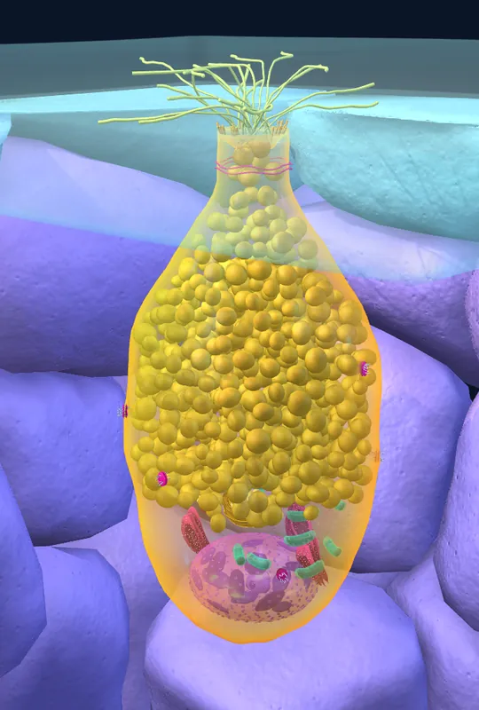 01MODEL
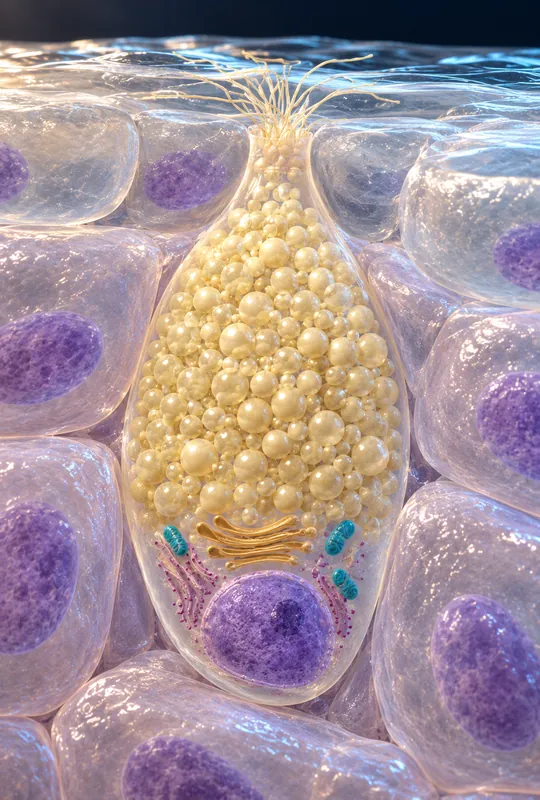 02AI STILL
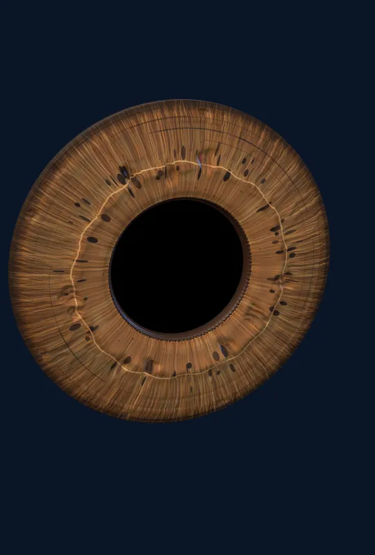 01MODEL
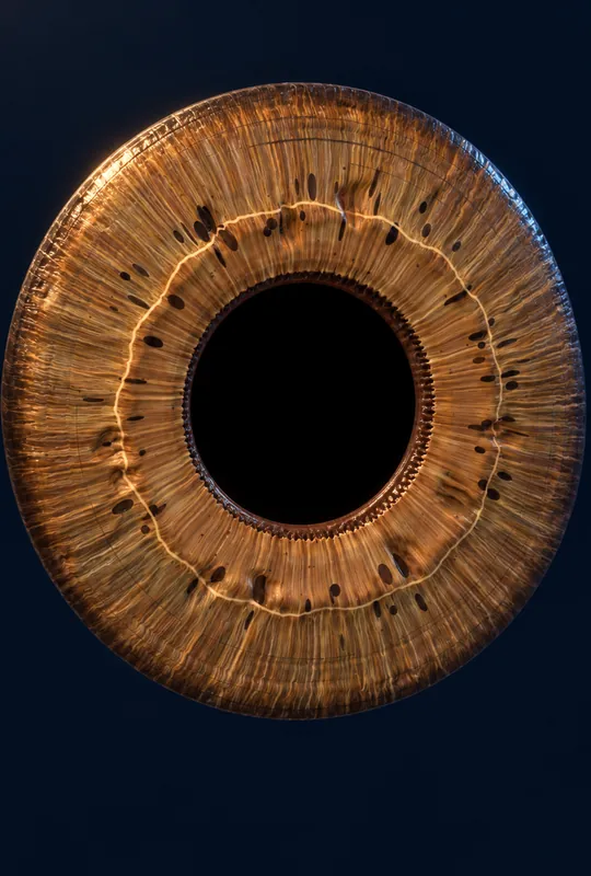 02AI STILL
AI expertise
AI Production PioneersHands-on experience with image and motion tools, turned into faster production at lower cost.
Image + motion enginesEfficiency
Lower Cost, Deeper ExpertiseAI speed and our team's expertise lower production costs while keeping accuracy intact.
Less cost · more outputHuman-led
Experts Directing AIHigher output, with human review and experts making the final call.
Final call: humanReferences
Referenced FactsEvery number backed by two to three independent sources.
2–3 sources per fact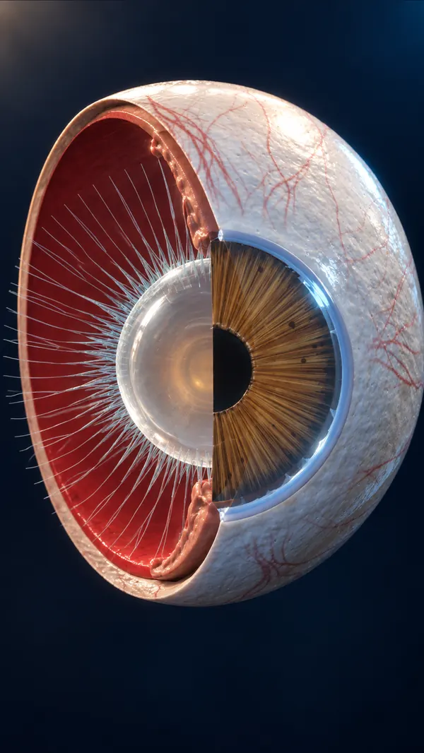
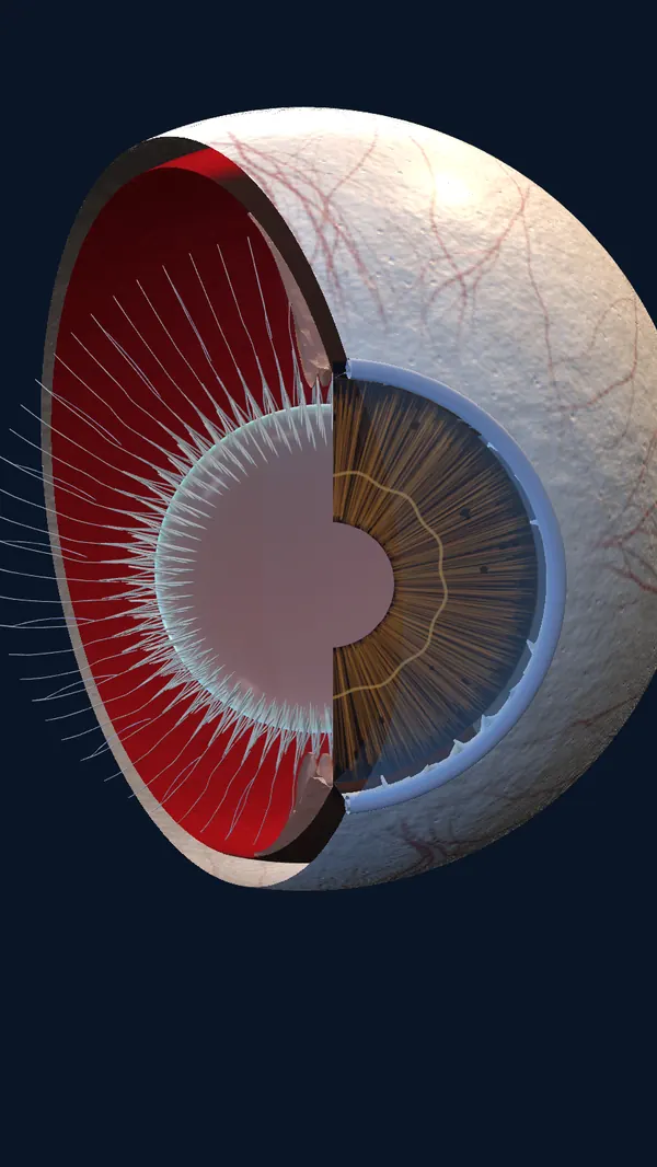 Atlas modelCinematic
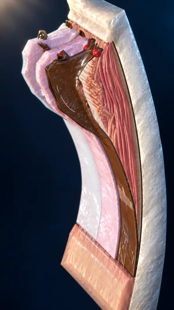
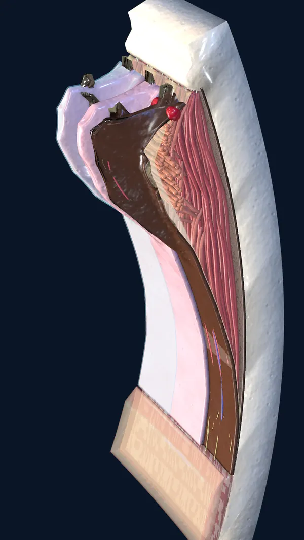 Atlas modelCinematic
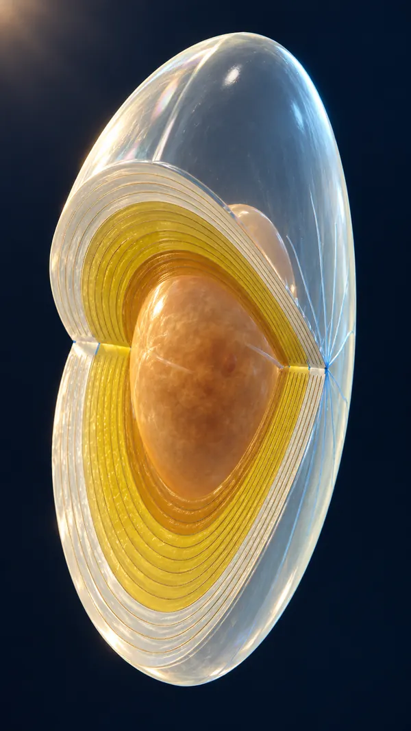
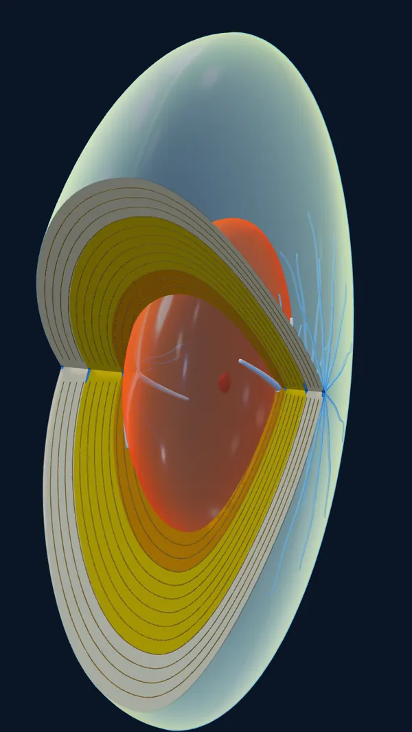 Atlas modelCinematic
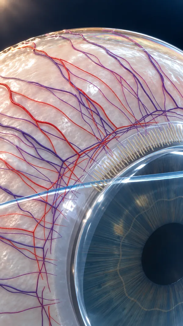
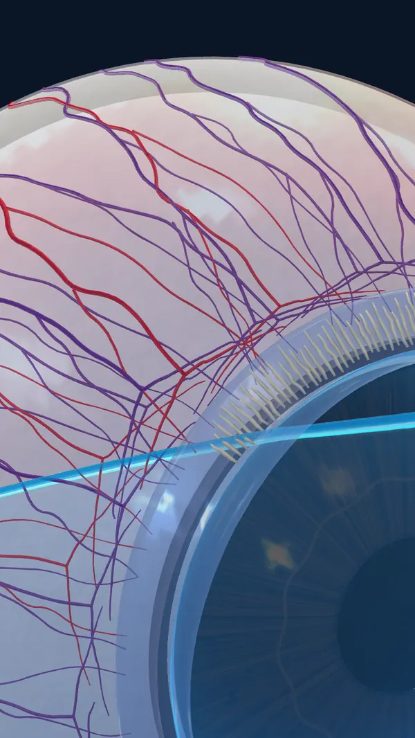 Atlas modelCinematic
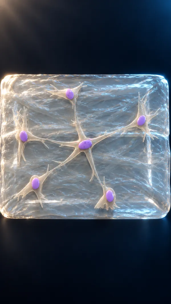
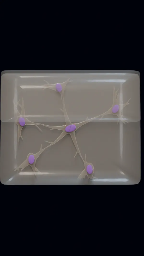 Atlas modelCinematic
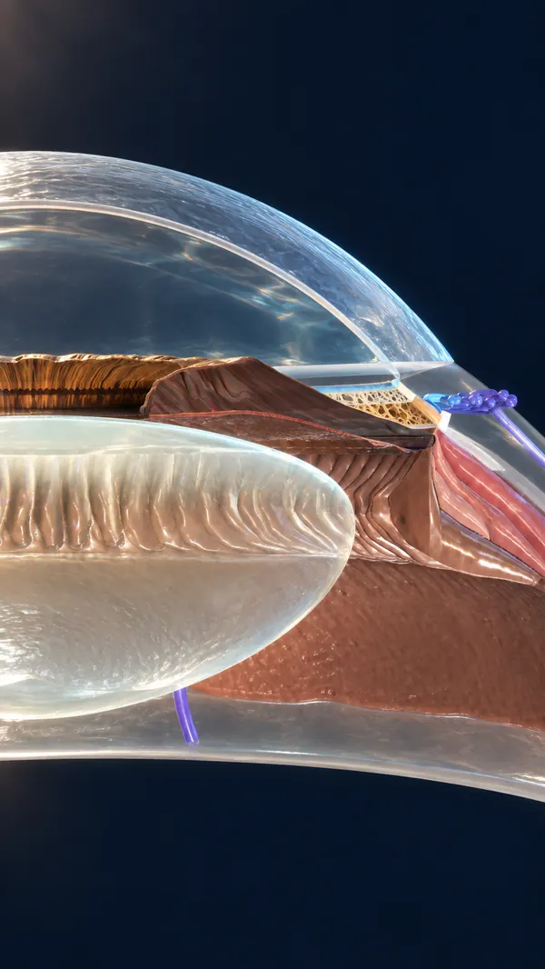
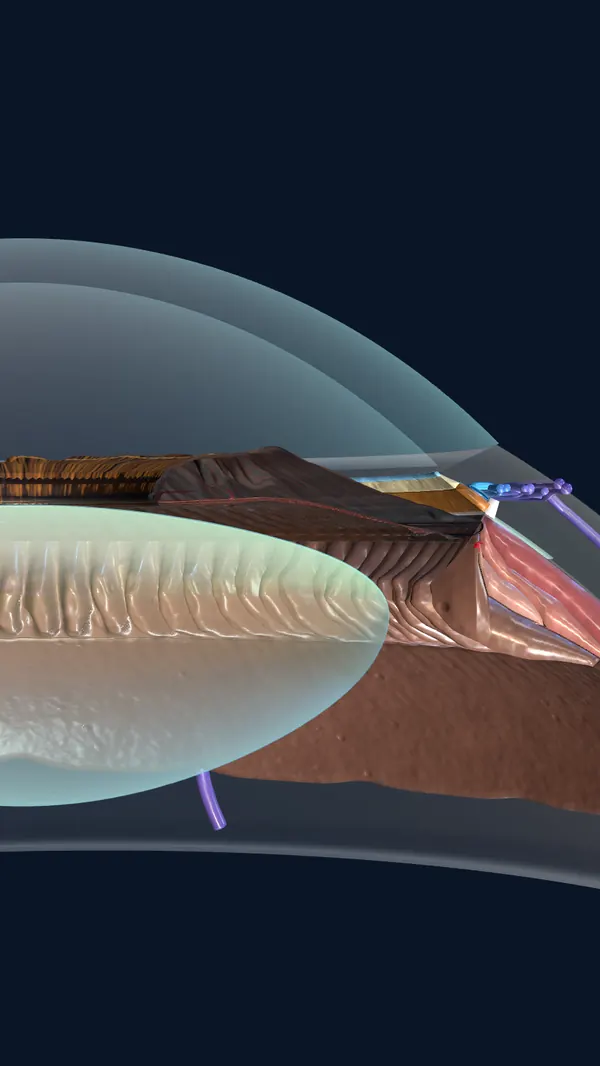 Atlas modelCinematic
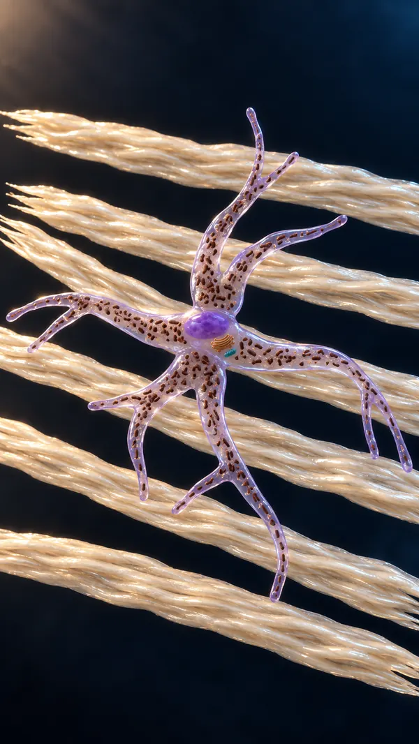
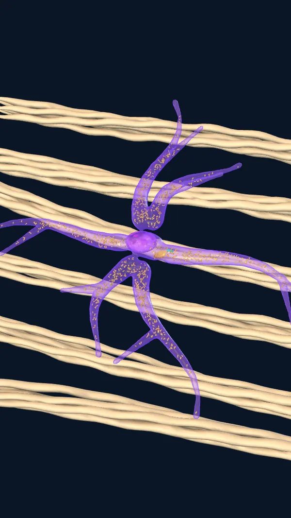 Atlas modelCinematic
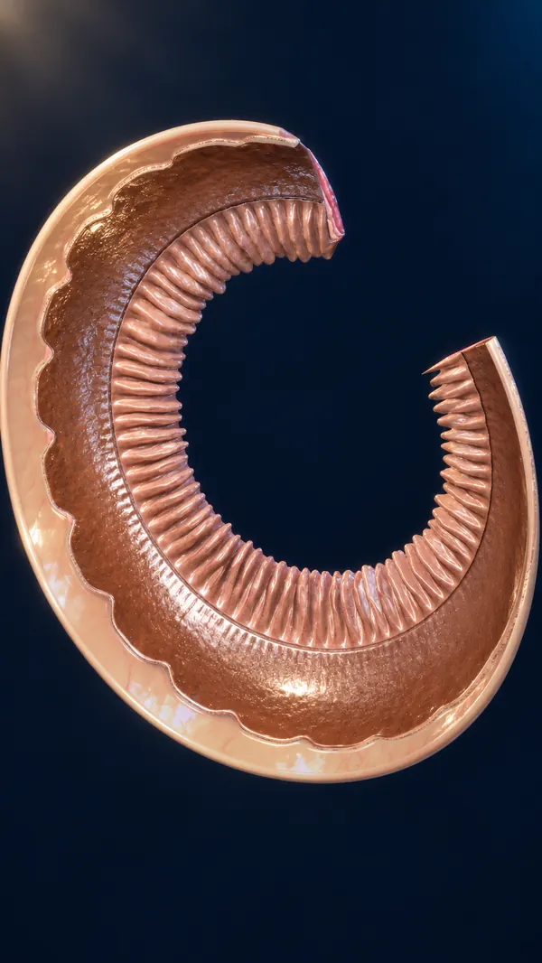
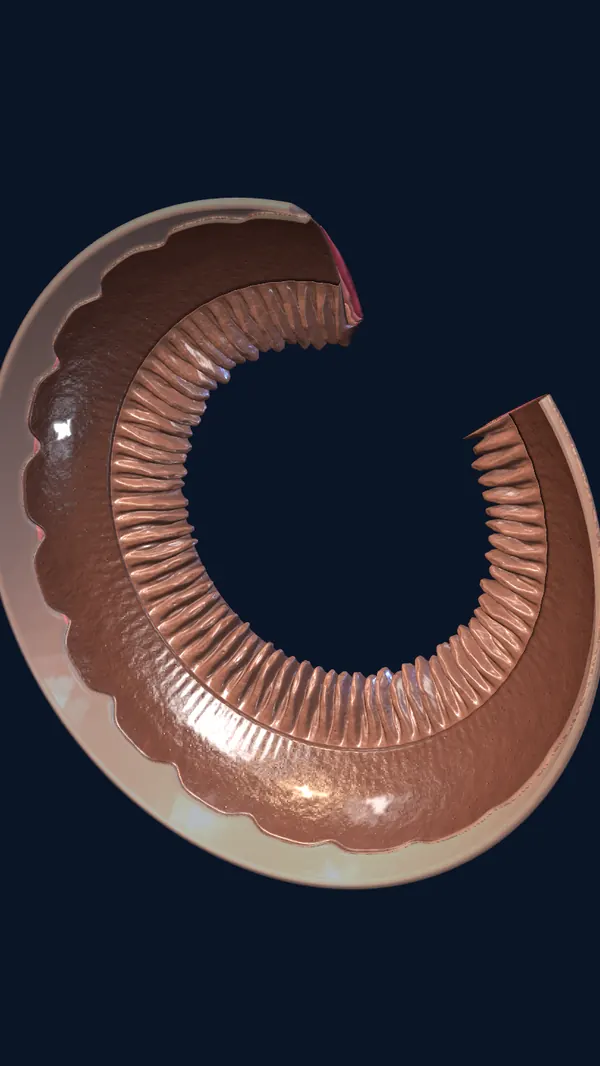 Atlas modelCinematic
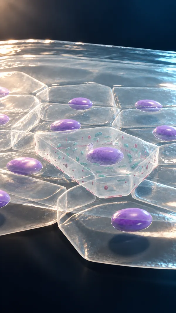
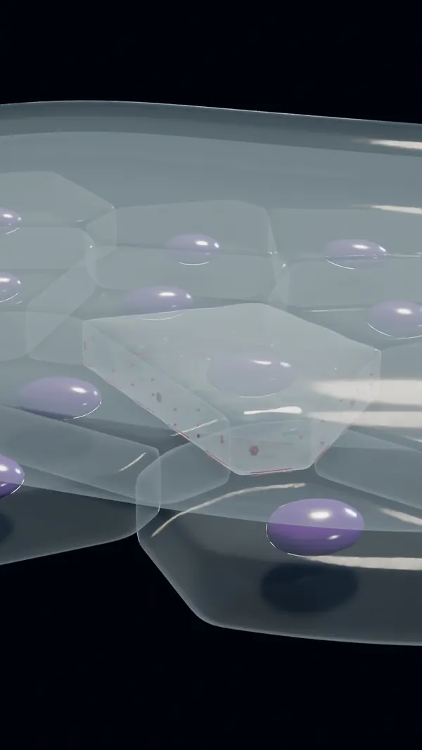 Atlas modelCinematic
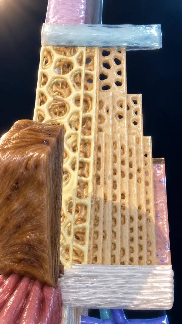
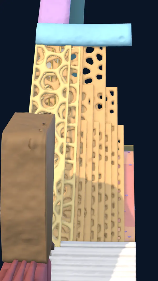 Atlas modelCinematic
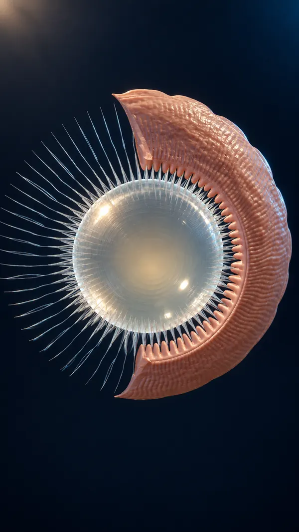
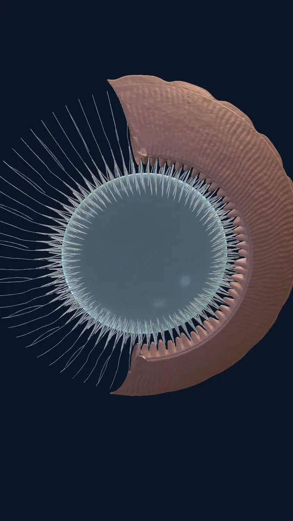 Atlas modelCinematic
 Atlas modelCinematic
 Atlas modelCinematic
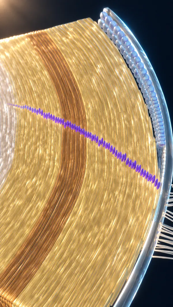
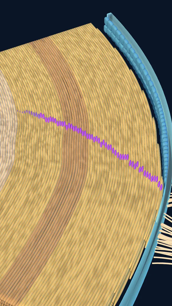 Atlas modelCinematic
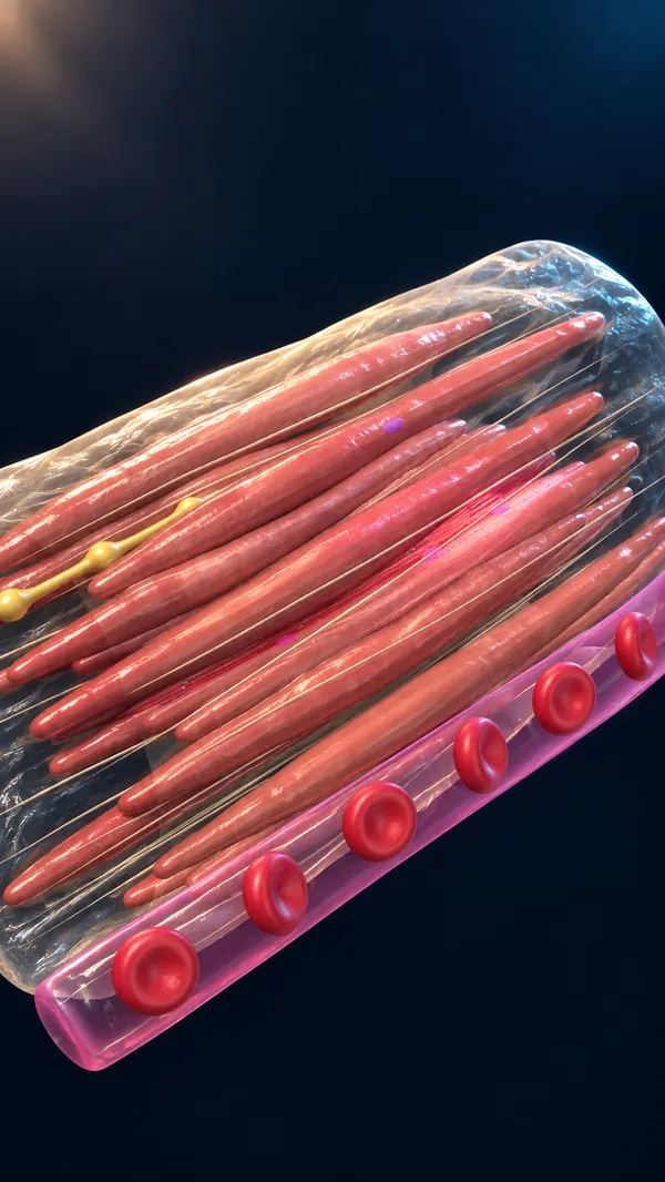
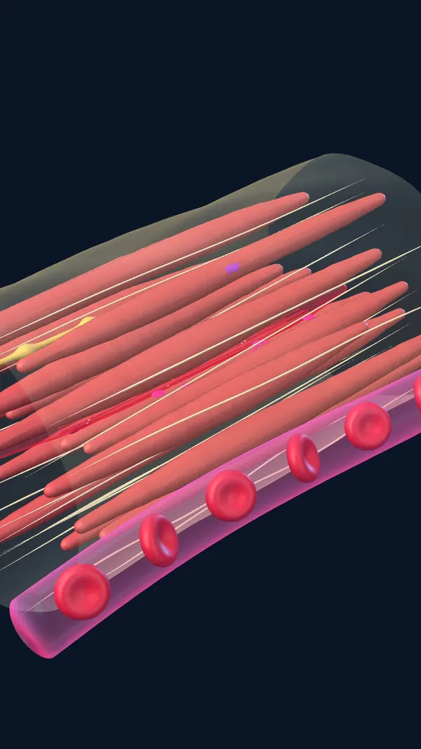 Atlas modelCinematic
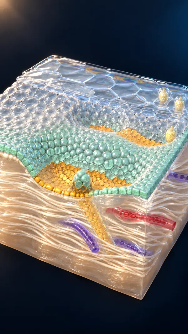
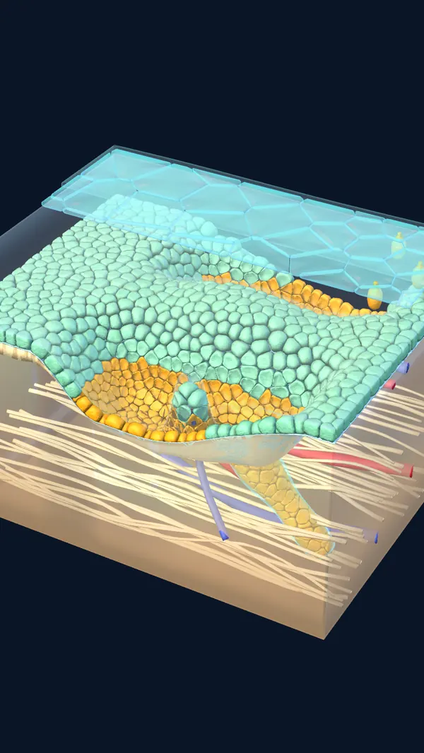 Atlas modelCinematic
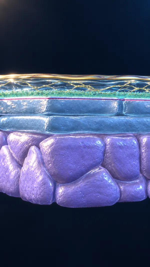
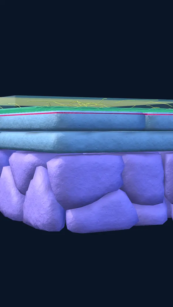 Atlas modelCinematic
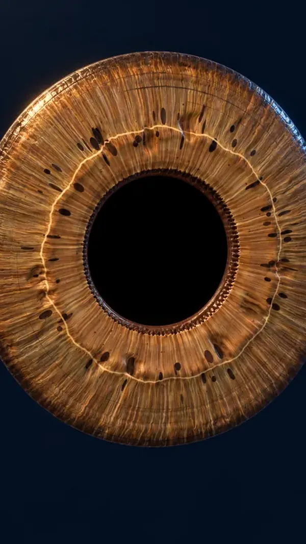
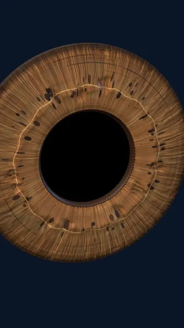 Atlas modelCinematic
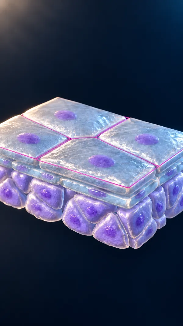
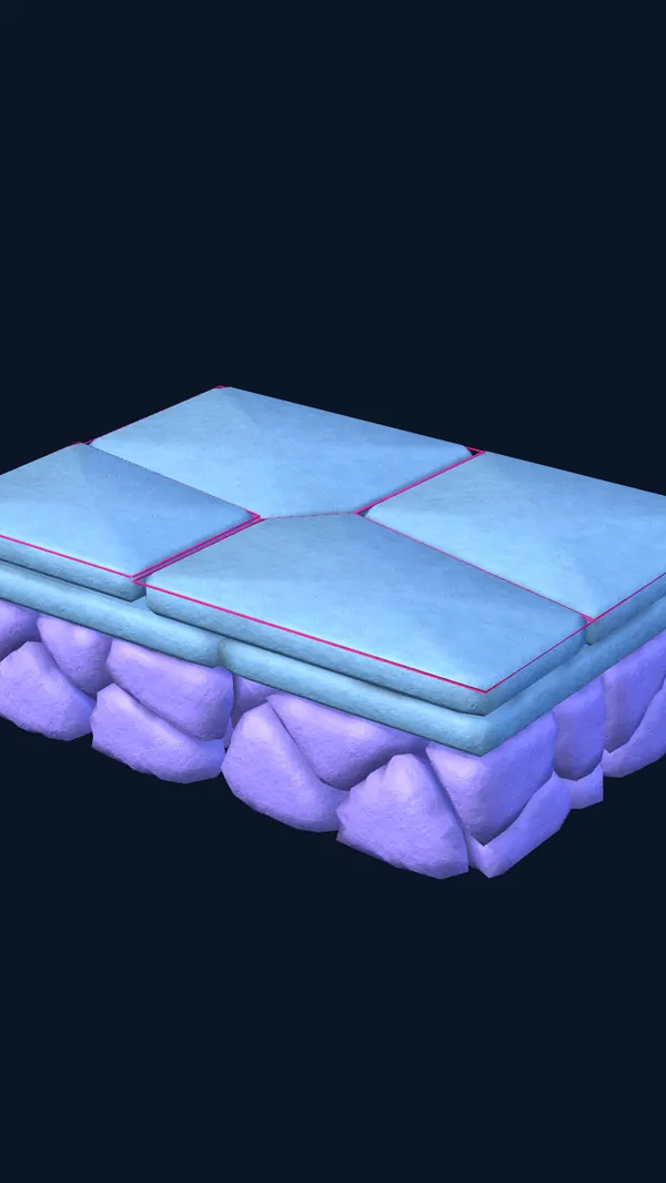 Atlas modelCinematic
 Atlas modelCinematic
 Atlas modelCinematic
 Atlas modelCinematic
And that's just the eye… Imagine the whole body.
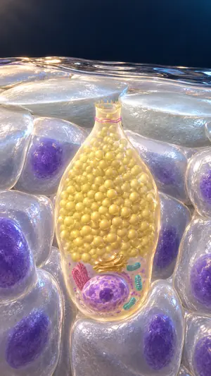 Ocular surface · cinematic image
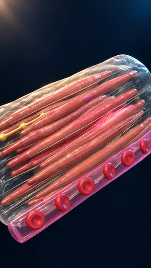 Iris · cinematic image
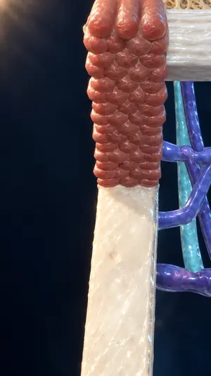 Aqueous outflow angle · cinematic image
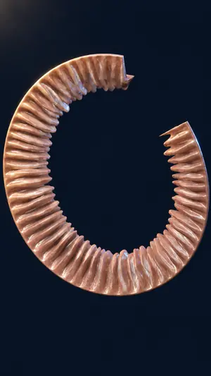 Ciliary body · cinematic image
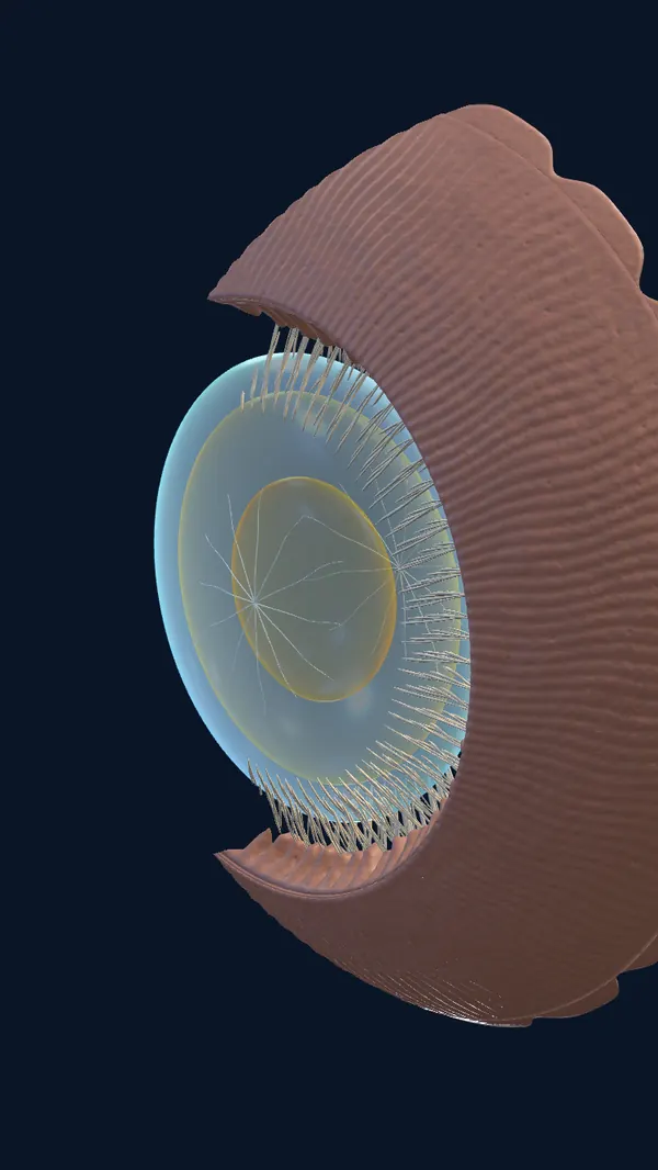
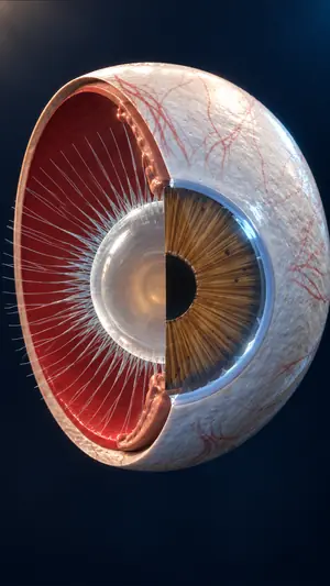 Lens and suspensory zonules · cinematic image
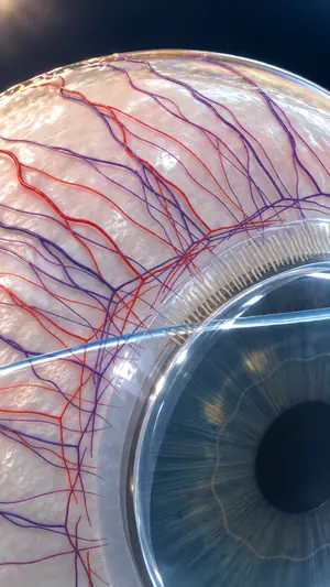 Ocular surface · cinematic image
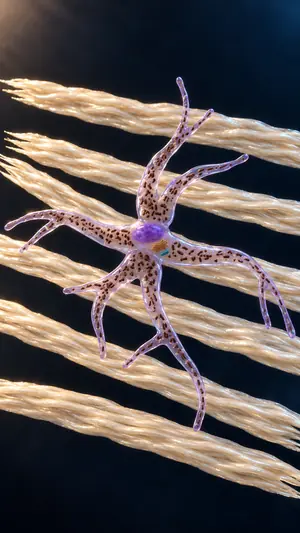 Iris · cinematic image
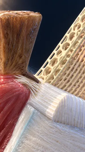 Aqueous outflow angle · cinematic image
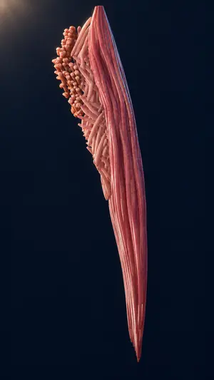 Ciliary body · cinematic image
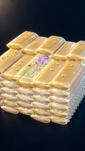 Lens and suspensory zonules · cinematic image
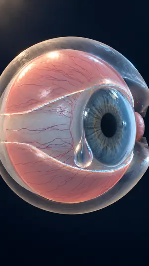 Ocular surface · cinematic image
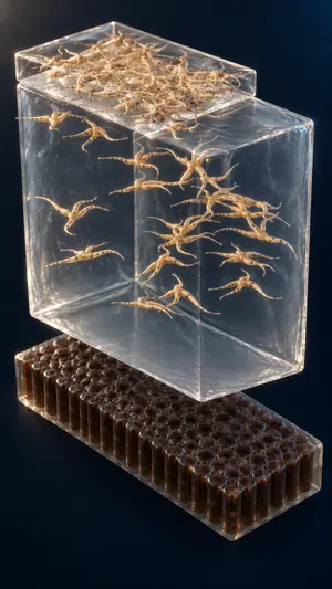 Iris · cinematic image
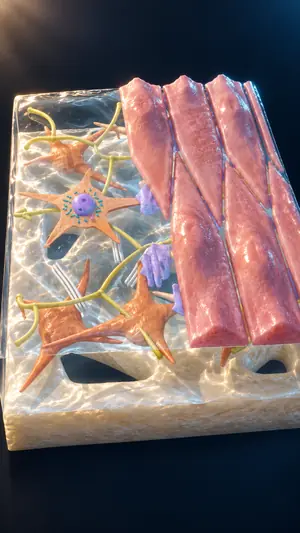 Aqueous outflow angle · cinematic image
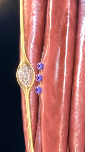 Ciliary body · cinematic image
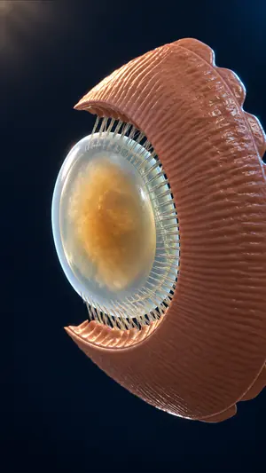 Lens and suspensory zonules · cinematic image
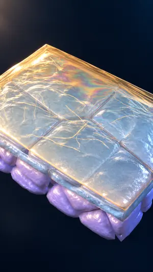 Ocular surface · cinematic image
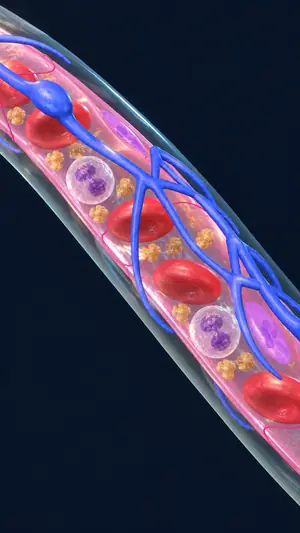 Iris · cinematic image
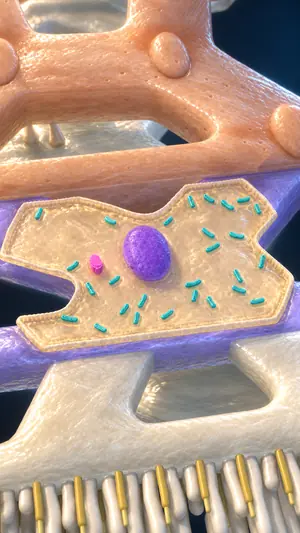 Aqueous outflow angle · cinematic image
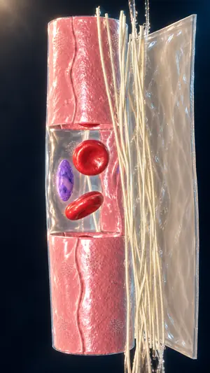 Ciliary body · cinematic image
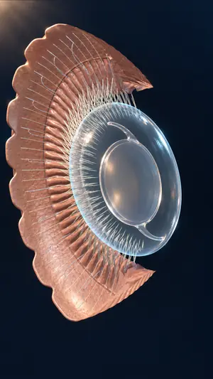 Lens and suspensory zonules · cinematic image
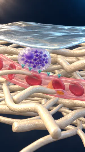 Ocular surface · cinematic image
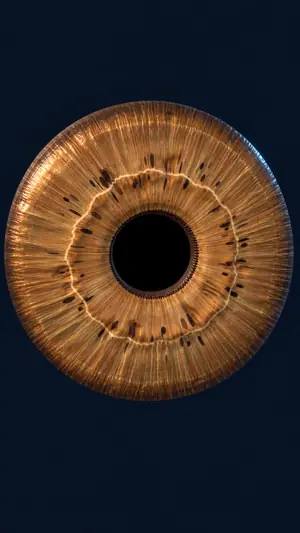 Iris · cinematic image
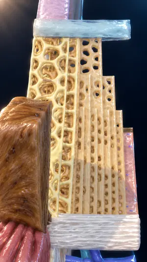 Aqueous outflow angle · cinematic image
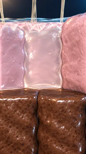 Ciliary body · cinematic image
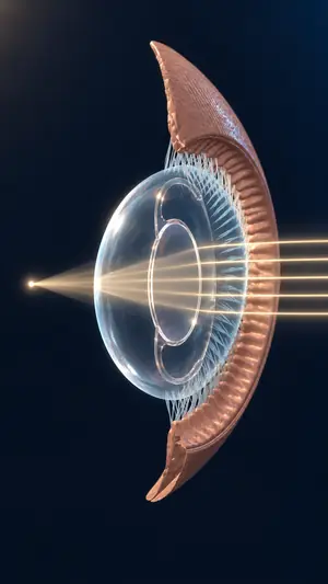 Lens and suspensory zonules · cinematic image
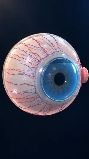 Ocular surface · cinematic image
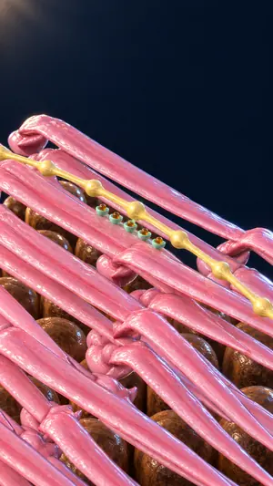 Iris · cinematic image
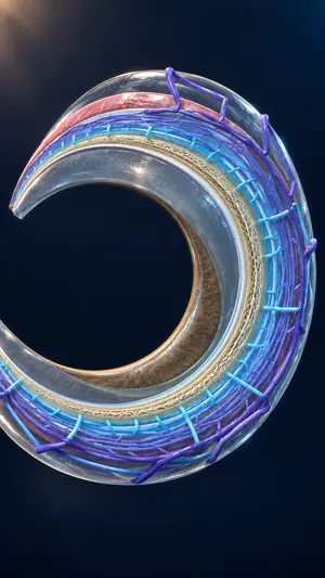 Aqueous outflow angle · cinematic image
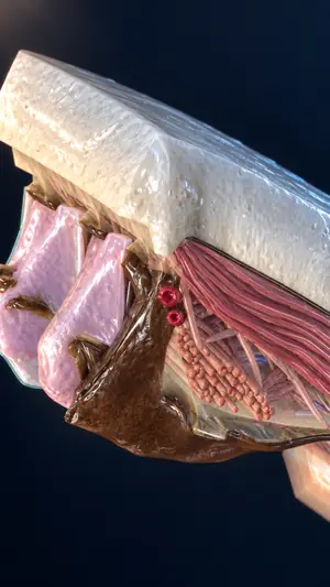 Ciliary body · cinematic image
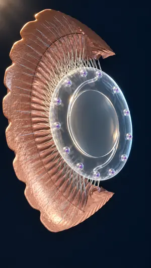 Lens and suspensory zonules · cinematic image
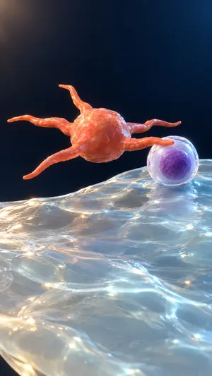 Ocular surface · cinematic image
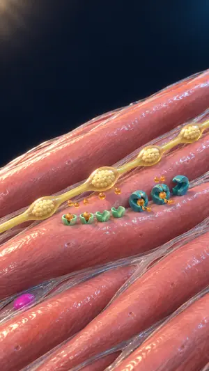 Iris · cinematic image
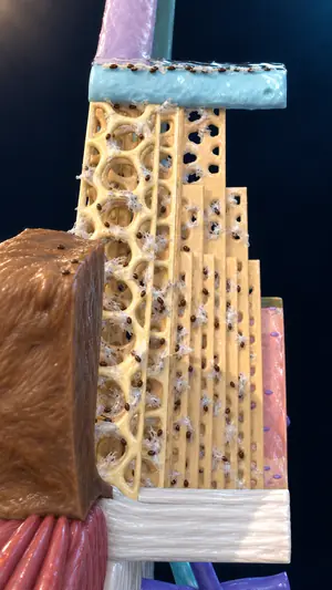 Aqueous outflow angle · cinematic image
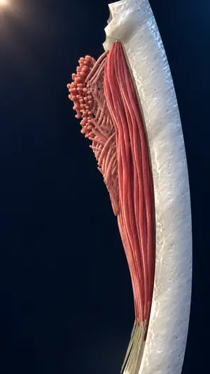 Ciliary body · cinematic image
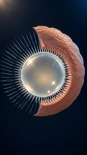 Lens and suspensory zonules · cinematic image
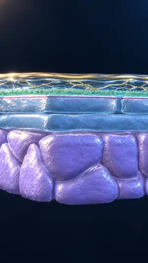 Ocular surface · cinematic image
 Iris · cinematic image
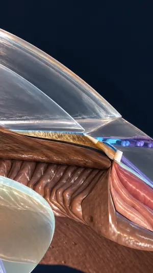 Aqueous outflow angle · cinematic image
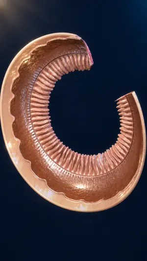 Ciliary body · cinematic image
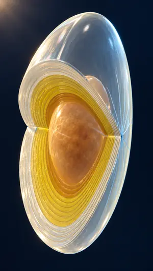 Lens and suspensory zonules · cinematic image
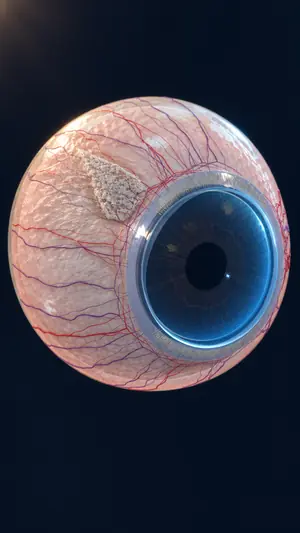 Ocular surface · cinematic image
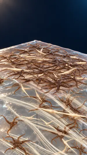 Iris · cinematic image
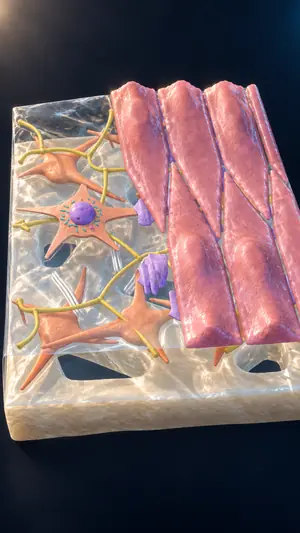 Aqueous outflow angle · cinematic image
 Ciliary body · cinematic image
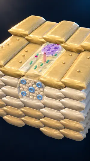 Lens and suspensory zonules · cinematic image
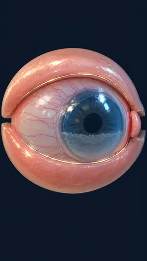 Ocular surface · cinematic image
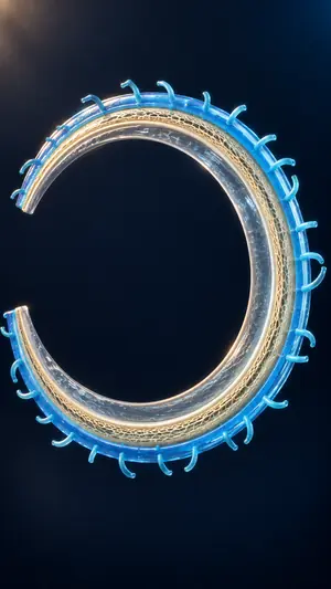 Aqueous outflow angle · cinematic image
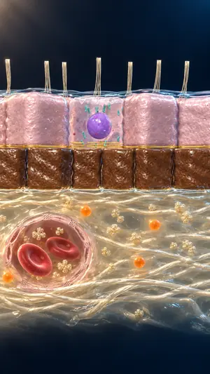 Ciliary body · cinematic image
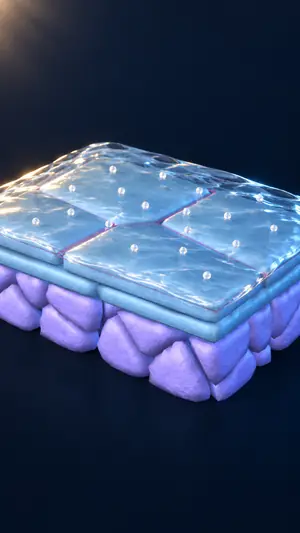 Ocular surface · cinematic image
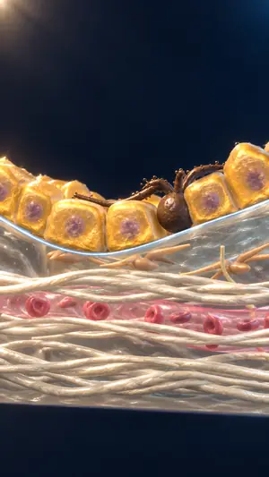 Ocular surface · cinematic image
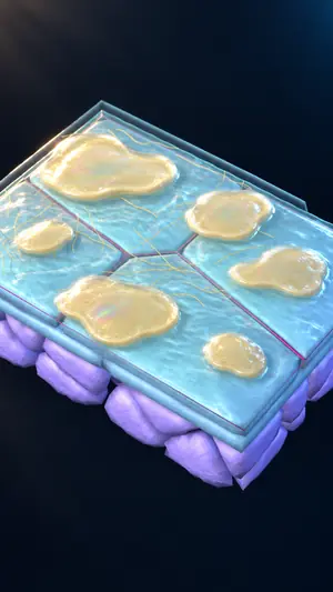 Ocular surface · cinematic image
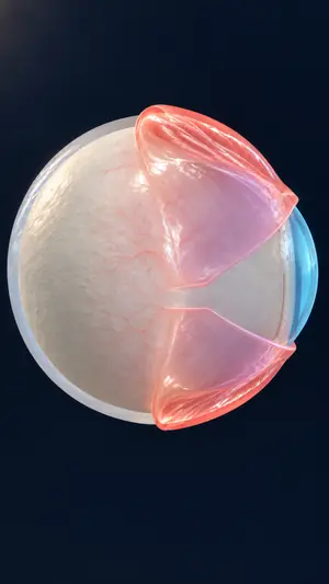 Ocular surface · cinematic image
Content that engages your audience with the same scientific structure, from our original models, with full copyright owned by Visualisy and a licence for your use.
For institutions
BenefitContent that serves your organisation's goal: sales and marketing, training and education, research, or awareness.
Present a drug or disease mechanism down to cell and molecule, as a video or an interactive model, for conferences, physician education and sales-team training.
Show your device and how it works inside organ and tissue, for training, sales presentations and conferences, with a custom model or one built on Visualisy models.
Organ, tissue, cell and molecule in one connected model, for lectures and e-learning in Arabic and English.
Turn your research mechanism or findings into a scientific visual, for a conference or a publication.
From the same models, we prepare content to train doctors and medical teams, and clear explanations for patients.
Add medical images and clips to your books, courses and platforms, under a commercial licence that defines the scope of use.
Content that serves your organisation's goal: sales and marketing, training and education, research, or awareness.
Choose what works for you
BenefitGet what you need in the form that fits your system, with usage rights clear from the start.
We prepare your app or interactive viewer, with installation and integration into your system through a technical partner.
Receive the models to integrate into your app or system, under a licence that fits your project.
Receive a video of a scientific mechanism, or images and clips for your presentation, publication or campaign.
Models remain Visualisy's property and you receive a licence to use them, unless a custom development contract states otherwise.
Scope your request with usResearch · build · review
We research the details and build the model, then review the science and the design. AI tools speed up the work, and experts review every stage.
BenefitFaster delivery, and a model with scientific references, reviewed by science and design specialists.
We compare counts, sizes and positions across two to three independent scientific sources.
We build each model on real scientific structure, with precise surface detail and soft lighting that shows it clearly.
We connect organ to tissue, then to cell, organelle and molecule.
Independent specialists review the science and design, then a doctor reviews the content.
Receive an interactive viewer, an app, a video or an image, as your project requires.
Speed
We build, review and improve in short cycles
Continuous releases: 49 app versions in about 3 months
Automated scene checks, frame by frame
Accuracy
We check every count, size and position against two to three independent sources
Independent scientific reviews, plus review by a consultant physician
Experts oversee every stage
Get your model faster, with referenced data, independent science and design reviews, then a doctor's review.
Selected from the Eye Atlas · © Visualisy
Click any image to see it at full size. © All models, images and videos are owned by Visualisy.
BenefitCompare the detail and display quality with what your project needs.
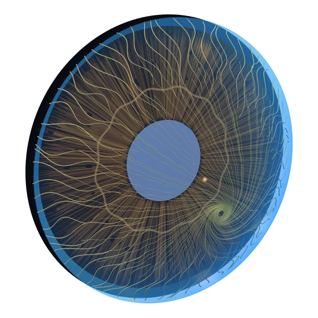
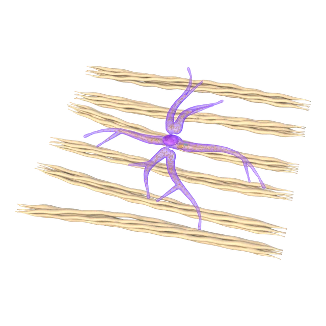
Cornea
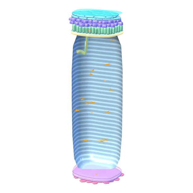
Corneal layers
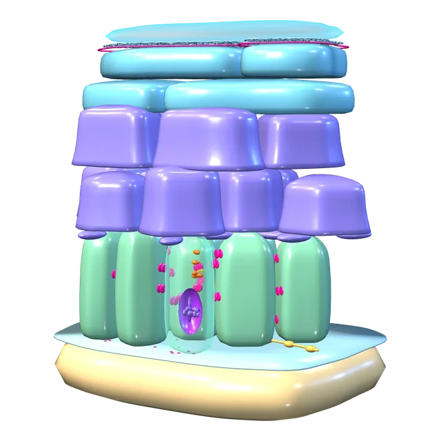
Corneal epithelial cells
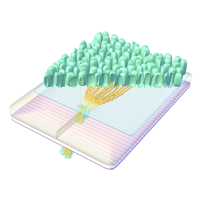
Corneal nerves
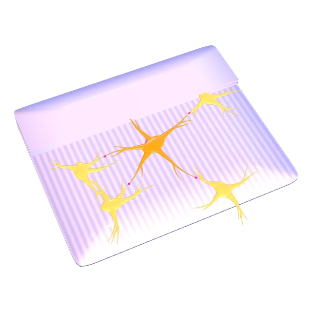
Keratocyte
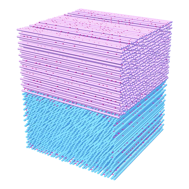
Collagen fibrils
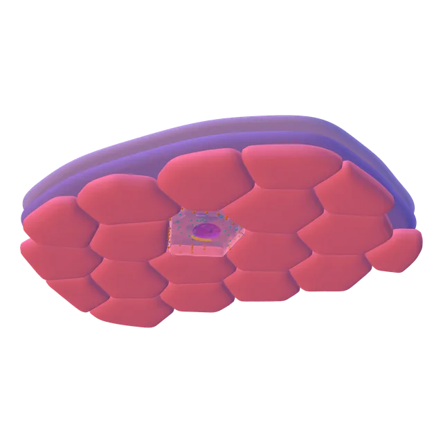
Corneal endothelial cells
Ocular surface
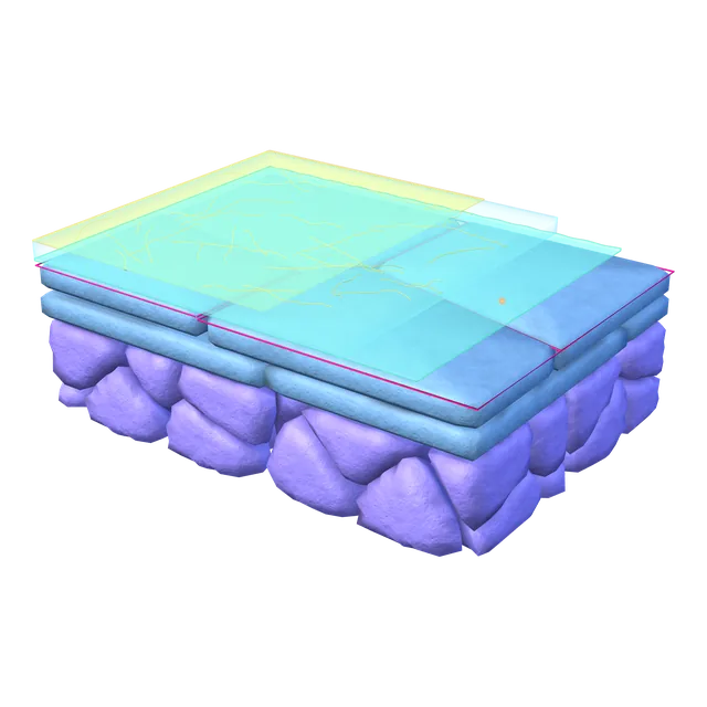
Tear film
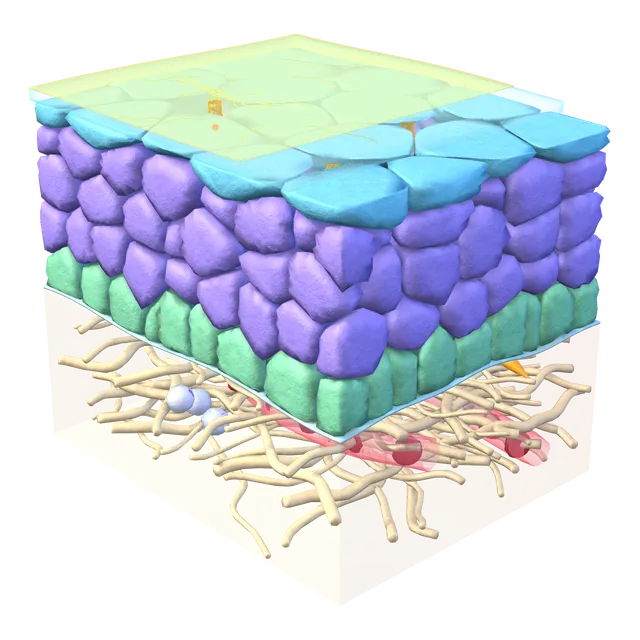
Conjunctival epithelium
Limbus
Goblet cell
Cell surface and glycocalyx
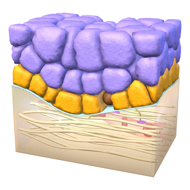
Stem cell niche
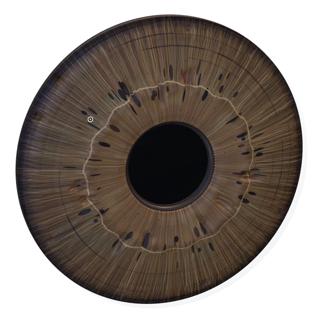
Iris and pupil
Iris layers
Sphincter muscle bundle
The two epithelial layers
Pigment cell
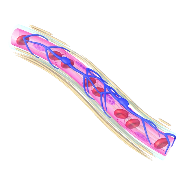
Iris capillary
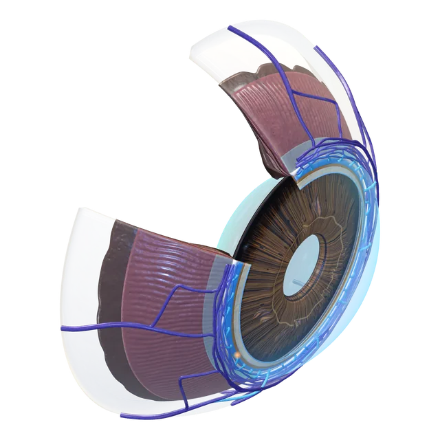
Drainage angle in the eye
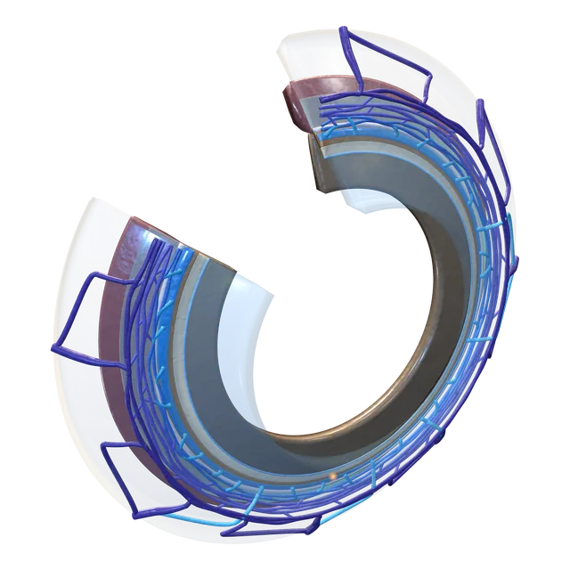
Drainage ring
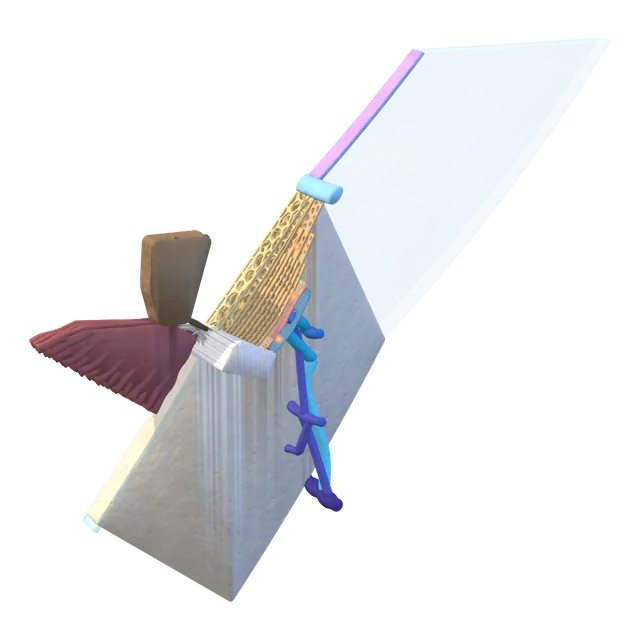
Drainage angle section
Trabecular beam
Juxtacanalicular tissue
Inner wall cells
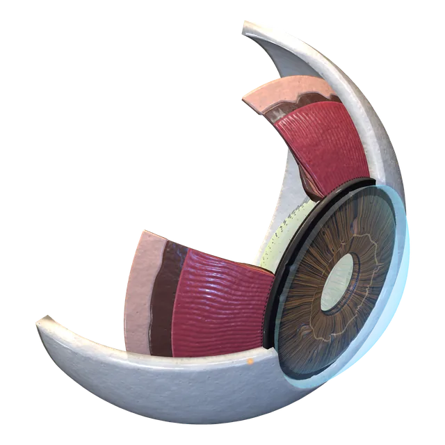
Ciliary body in the eye
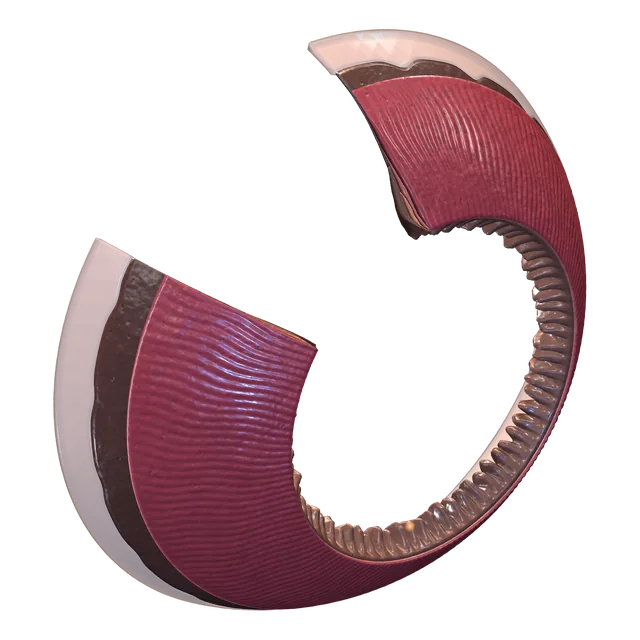
Ciliary body ring
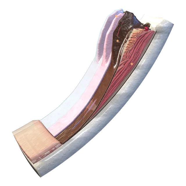
Histological section
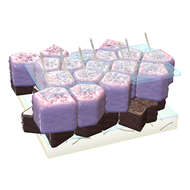
Bilayered epithelium
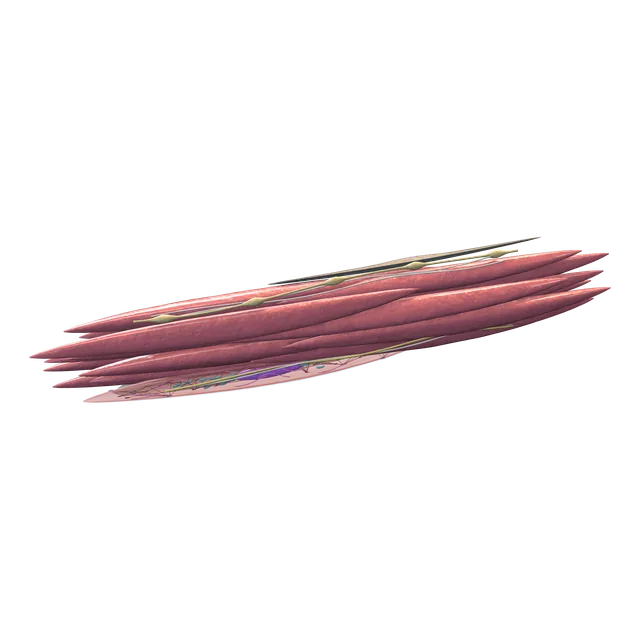
Ciliary muscle cells
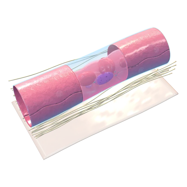
Fenestrated capillary
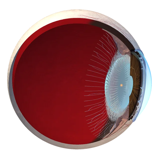
Lens in the eye
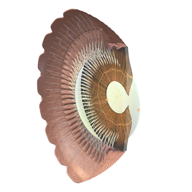
Lens and suspensory zonules
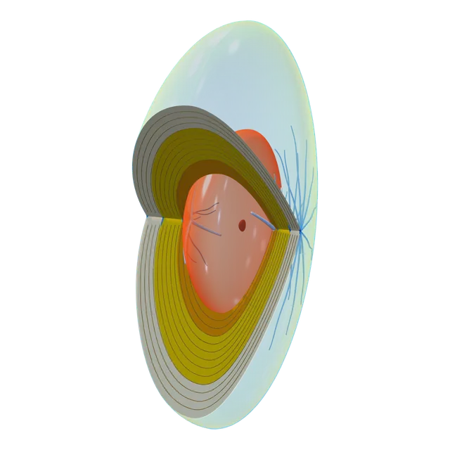
Nuclear zones and lens sutures
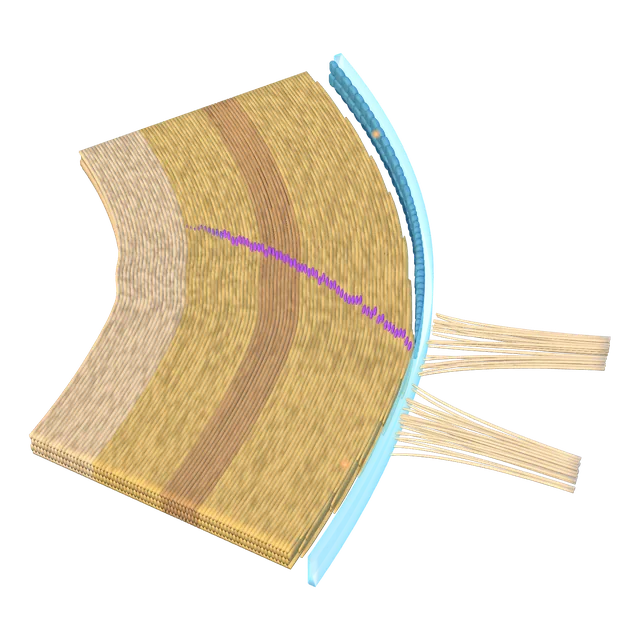
Equatorial lens bow
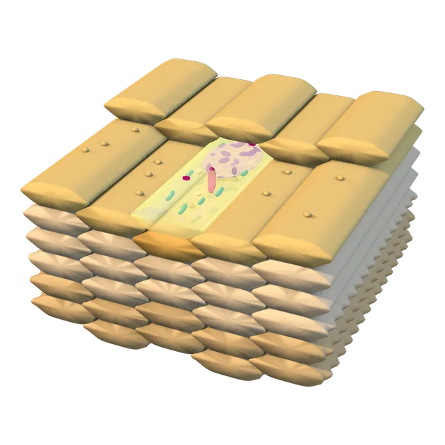
Lens fibres
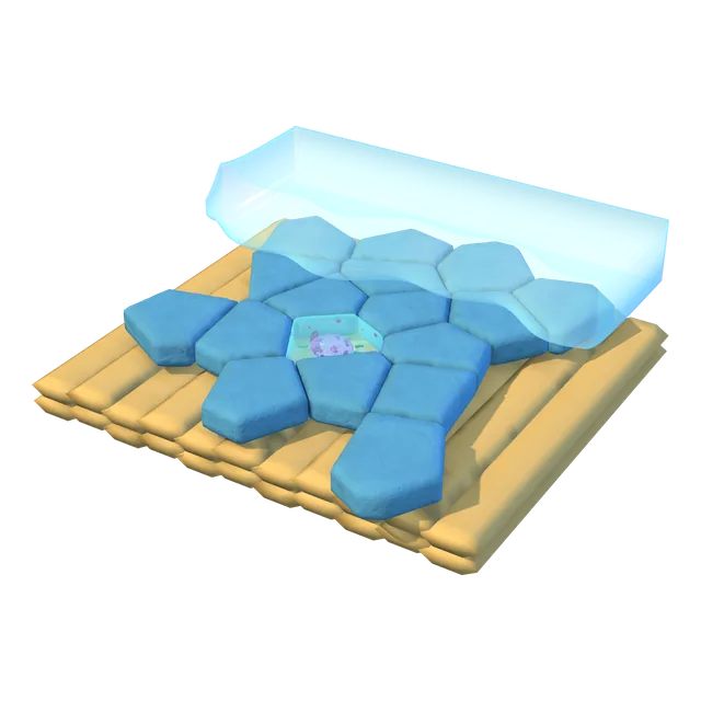
Corneal epithelial cells

Interwoven zonular fibres
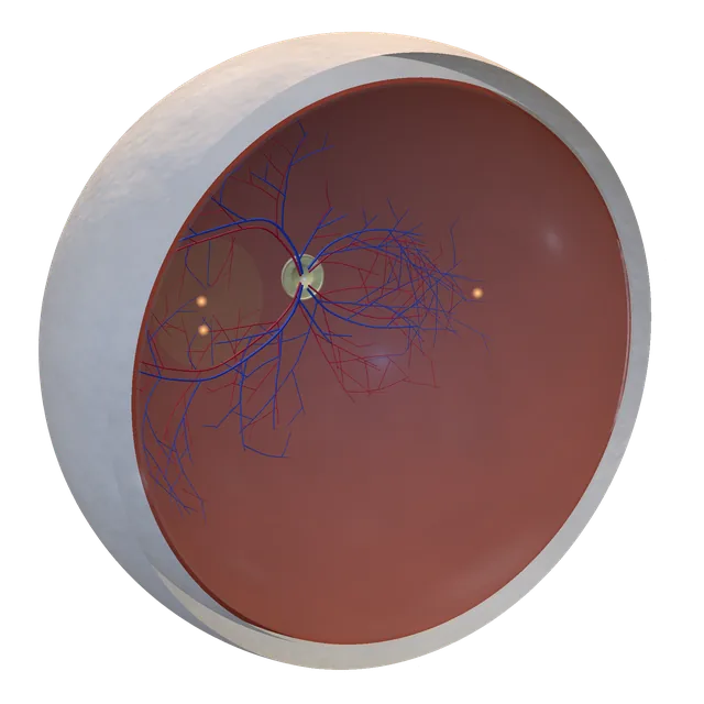
Retina in the eye
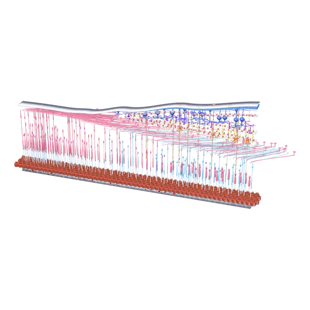
Foveal section
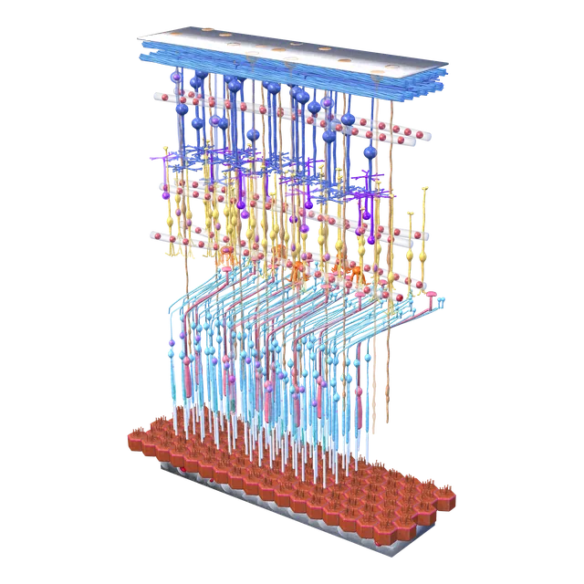
Parafoveal retinal layers
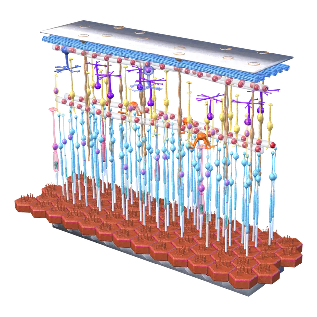
Peripheral retinal layers
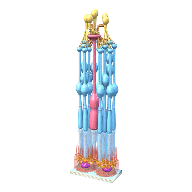
Photoreceptor cluster
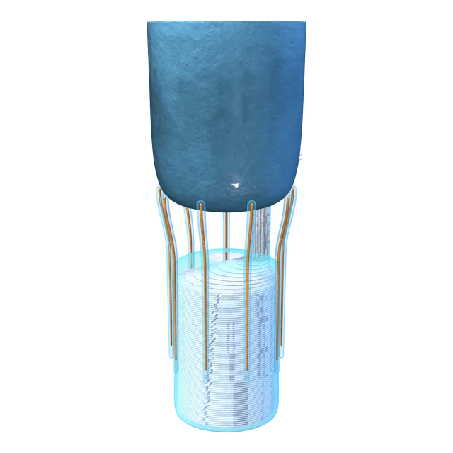
Outer segment base
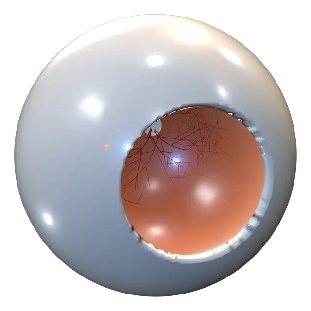
Retina in the eye
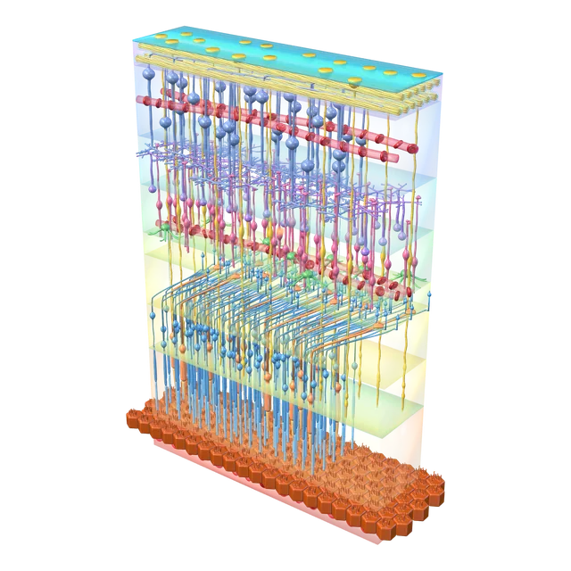
Parafoveal retinal layers
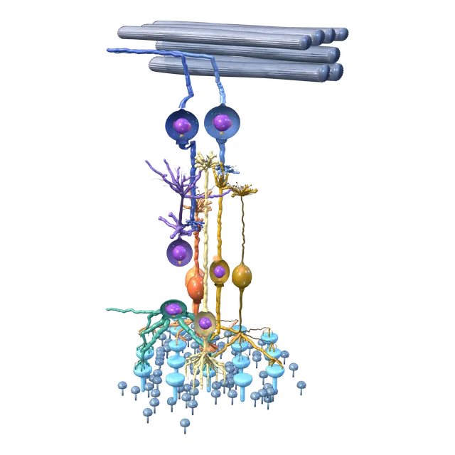
Inner retina column
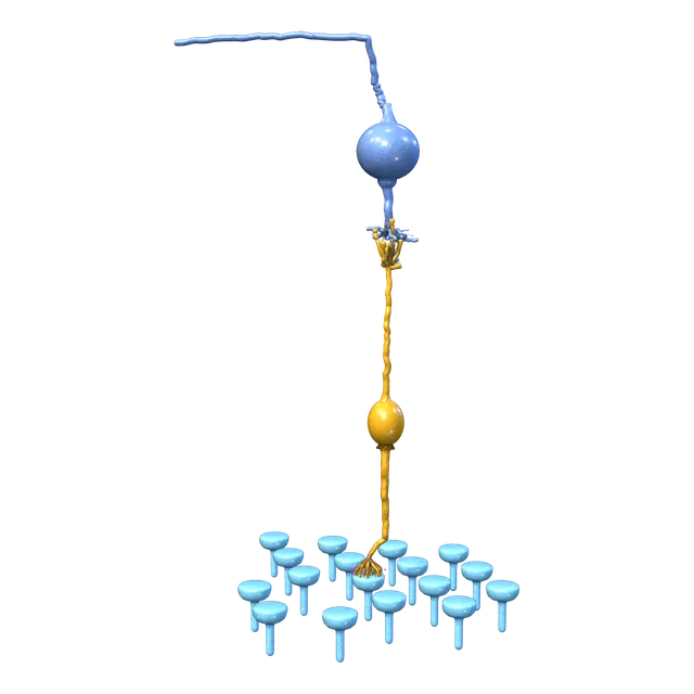
Vertical pathway, isolated
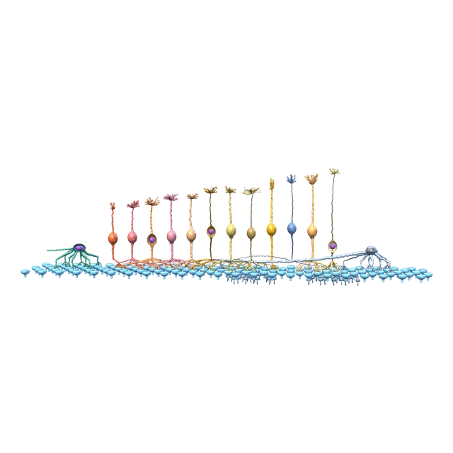
Bipolar cell types
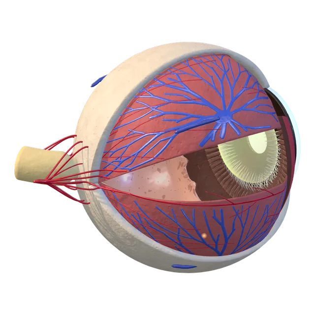
Choroid in the eye
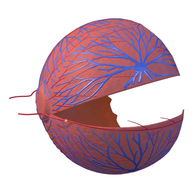
Choroid, isolated
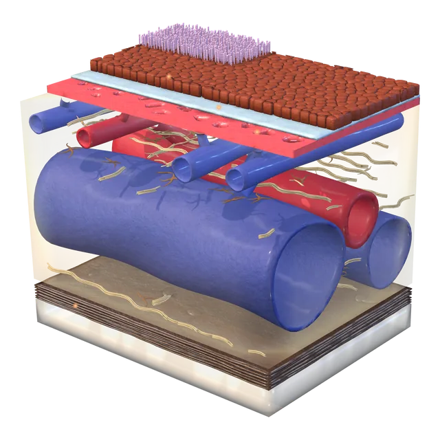
Tissue column
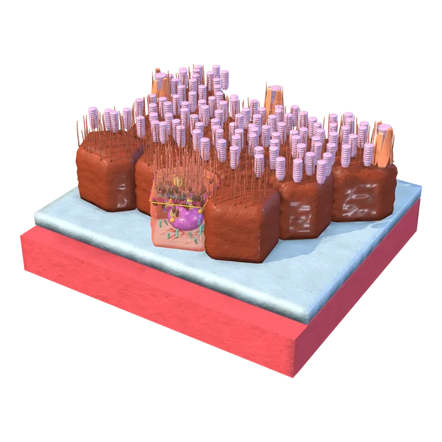
Retinal pigment epithelium (RPE) cells
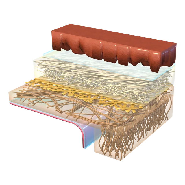
Bruch's membrane
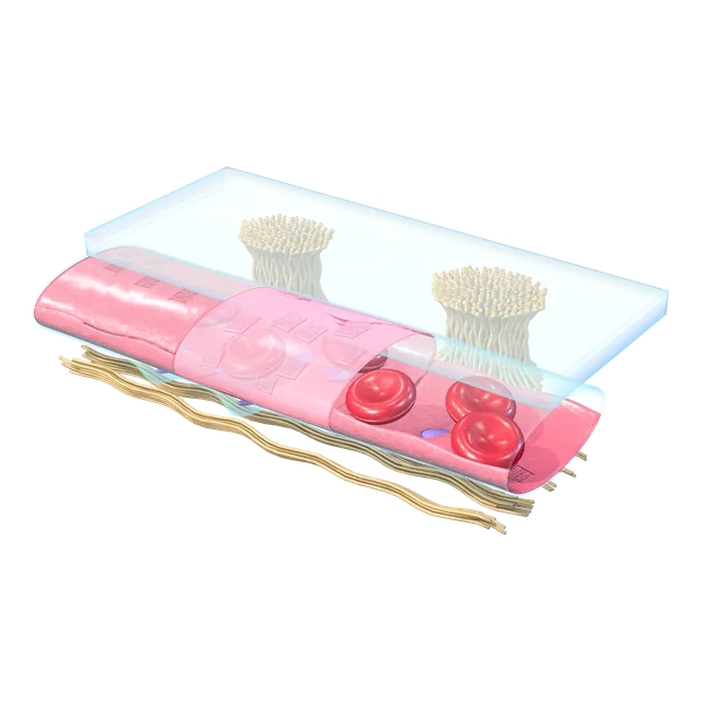
Choroidal capillary
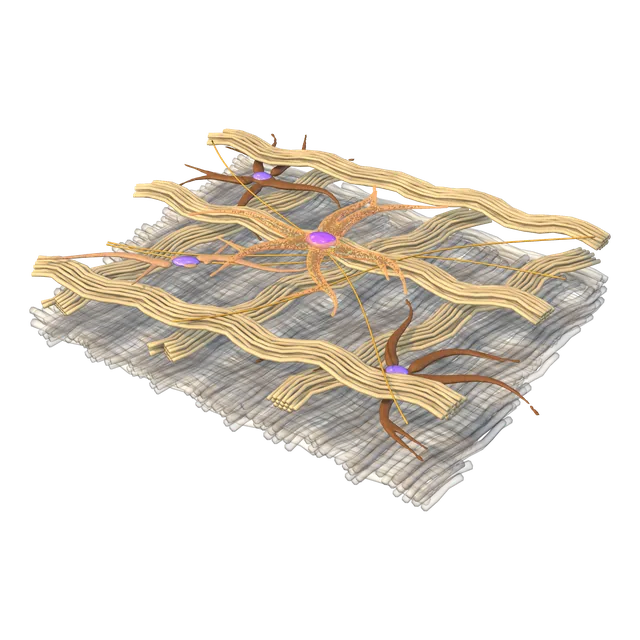
Melanocyte
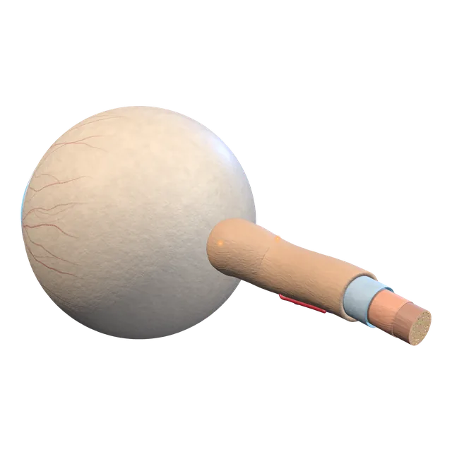
Optic nerve in the orbit
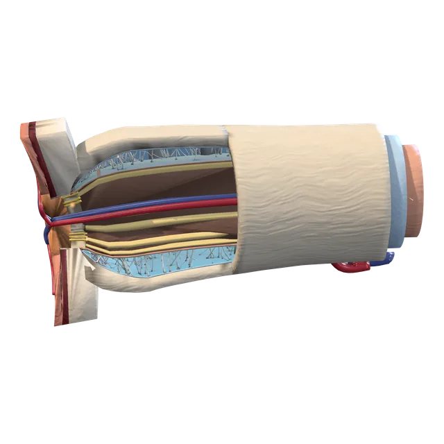
Optic nerve head
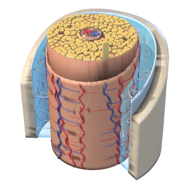
Nerve cross-section
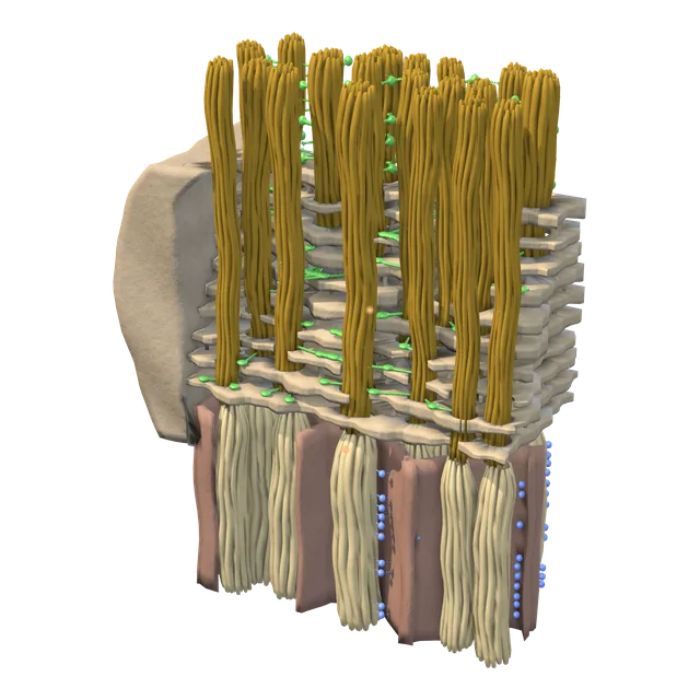
Lamina cribrosa
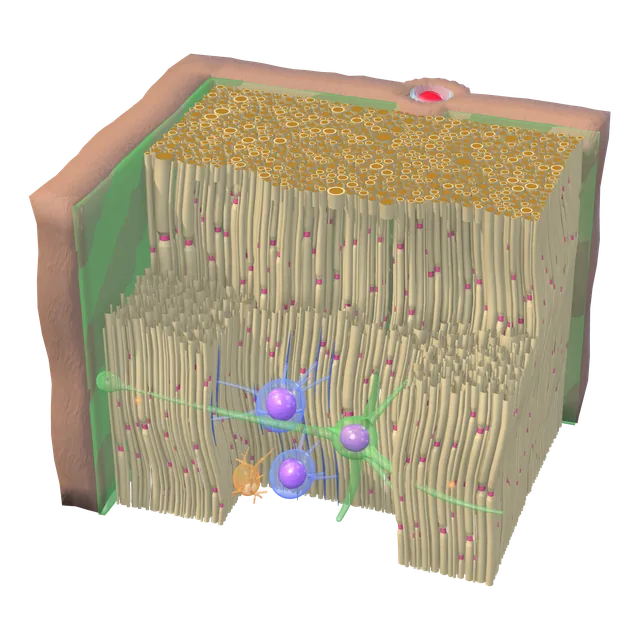
Inside a nerve fascicle
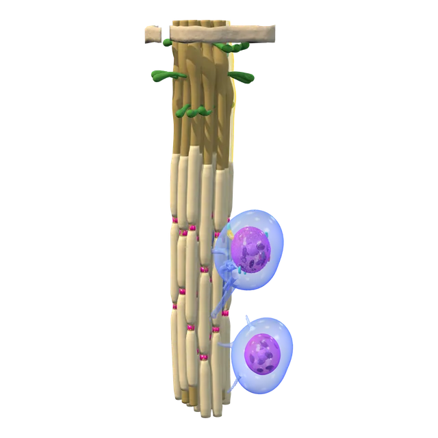
Axon and myelin
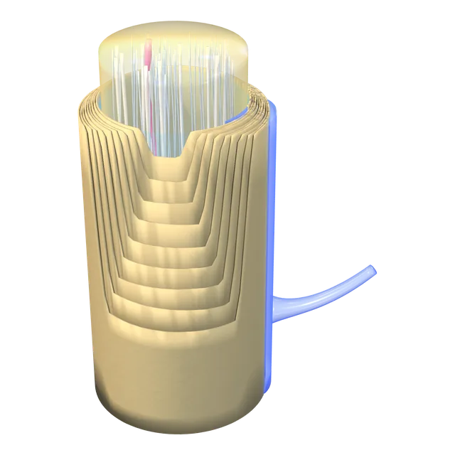
Myelin lamellae
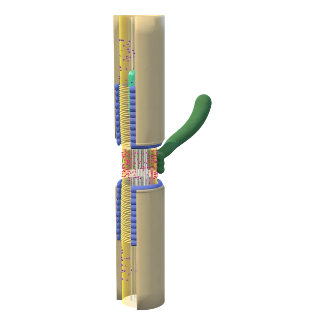
Node of Ranvier
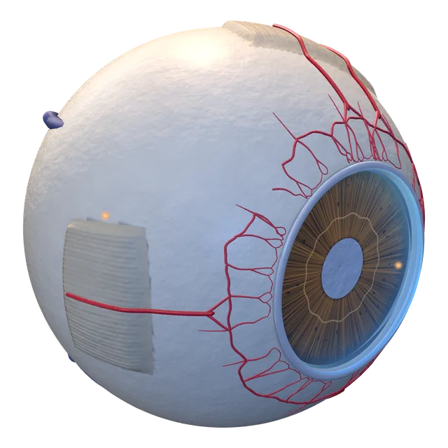
Sclera in the eyeball
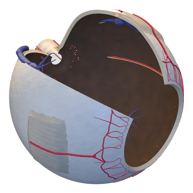
Sclera, isolated
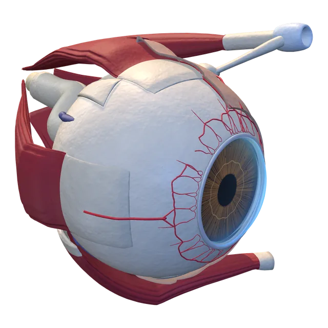
Tenon's capsule
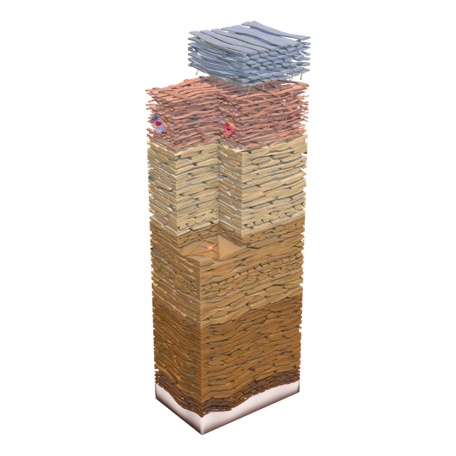
Scleral wall
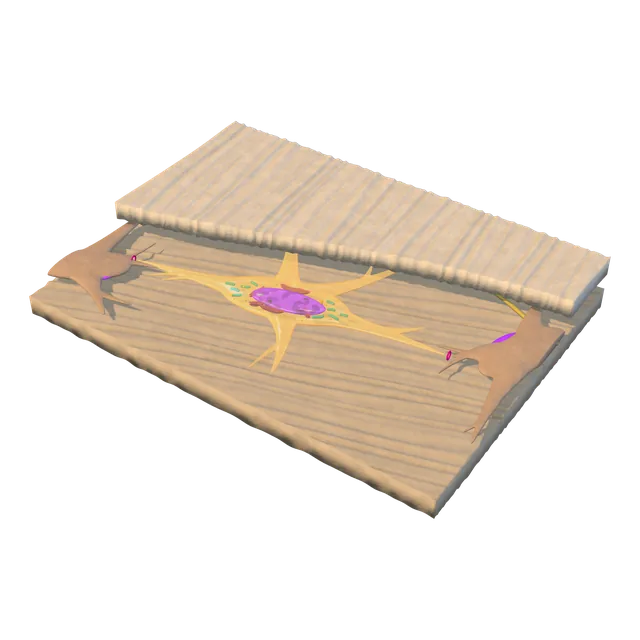
Fibroblast
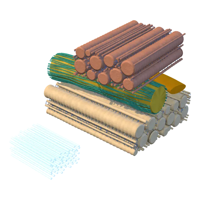
Collagen weave
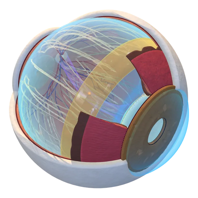
Vitreous in the eye
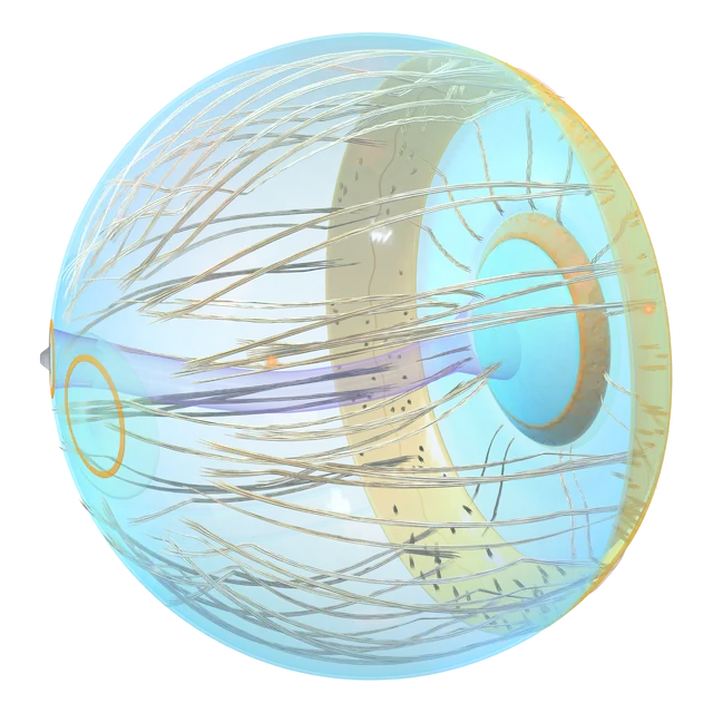
Vitreous, isolated
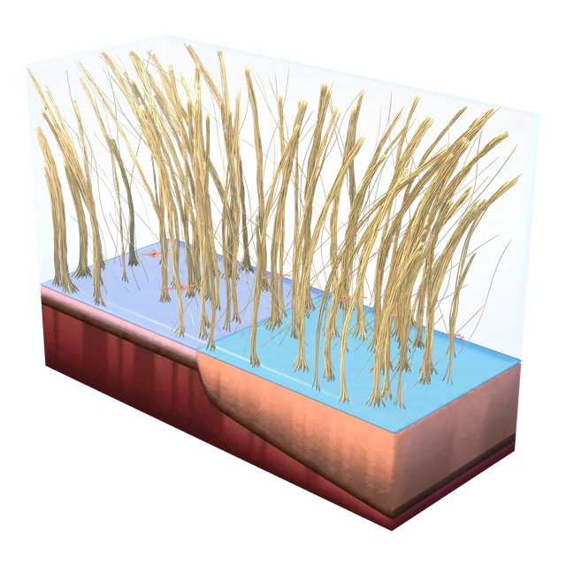
Vitreous base
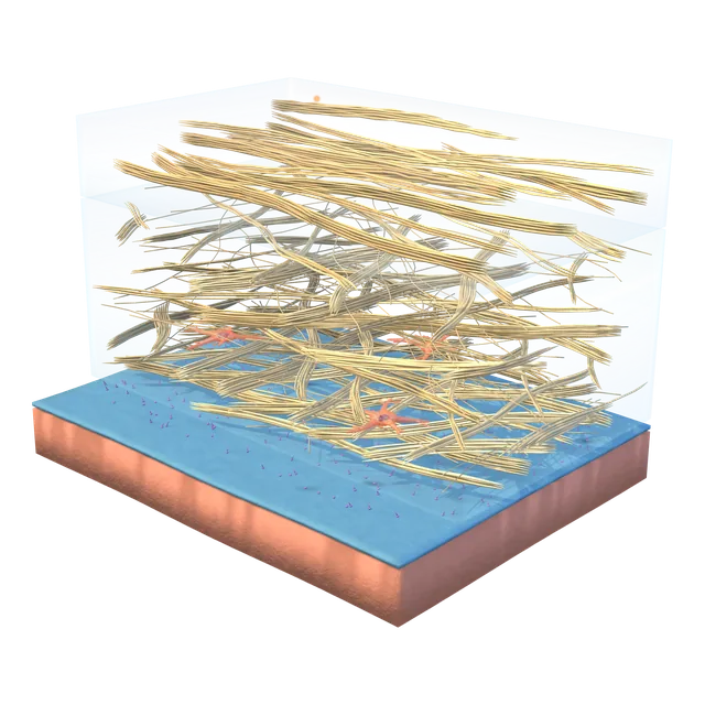
Posterior cortex and retinal surface
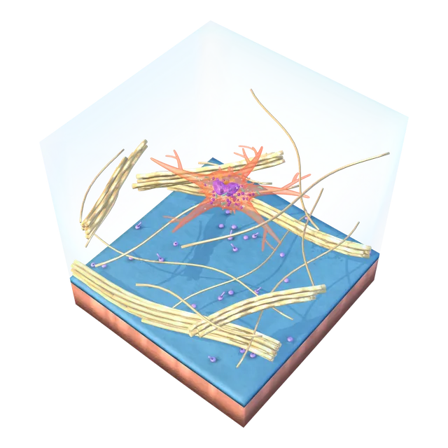
Hyalocyte
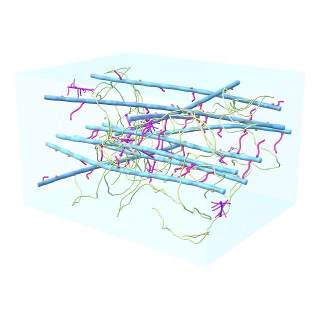
Collagen and hyaluronan network
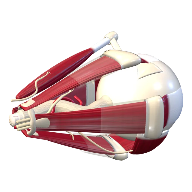
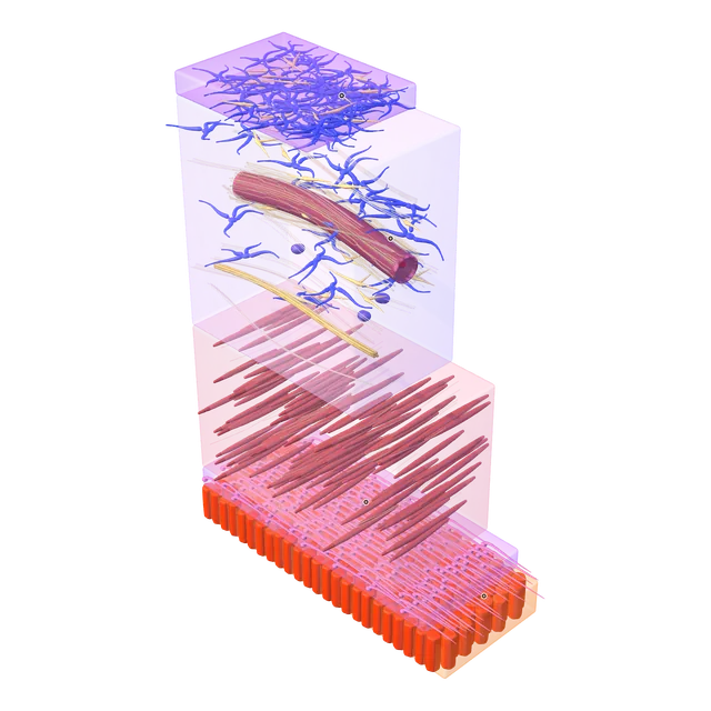
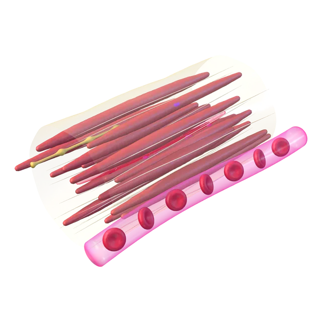
The eye and its six extraocular muscles
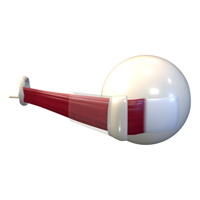
Lateral rectus
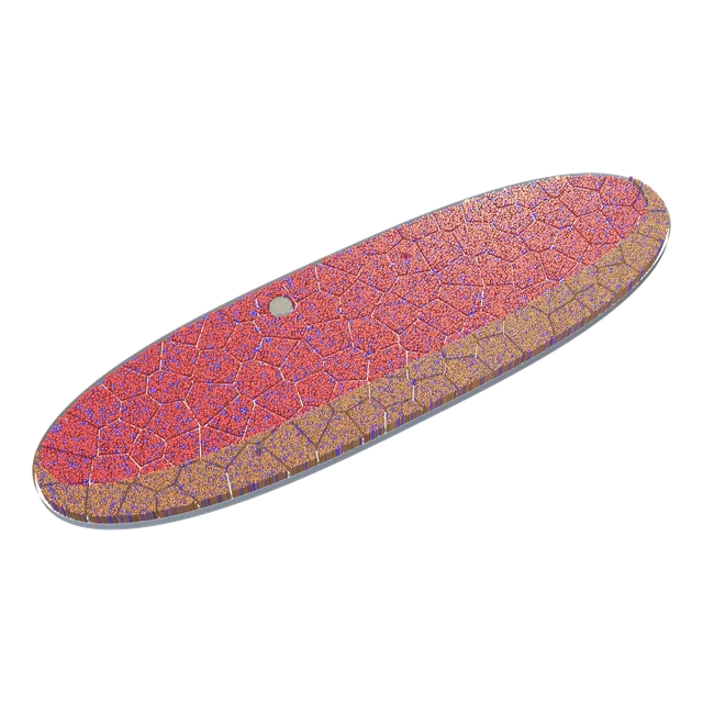
Muscle section
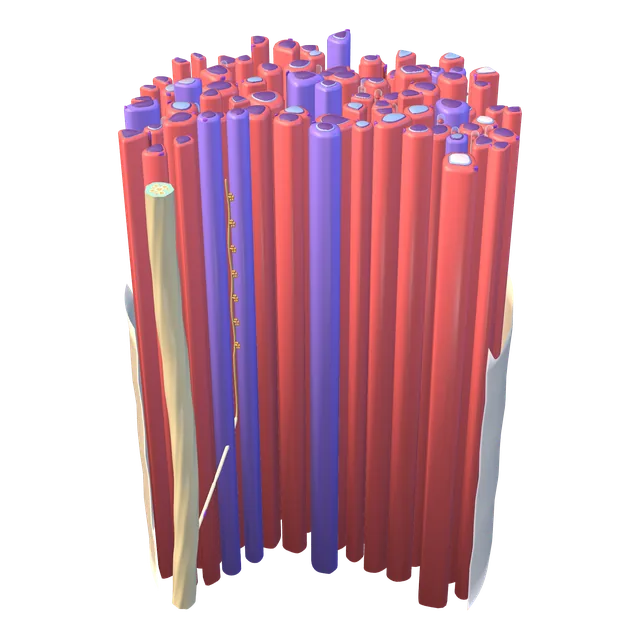
Muscle fibre bundle
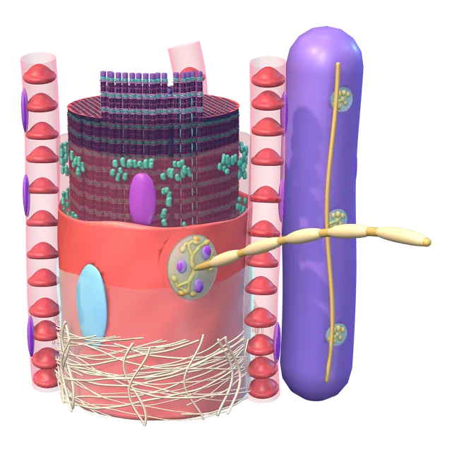
Muscle fibre
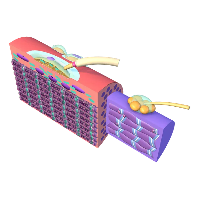
Neuromuscular junction
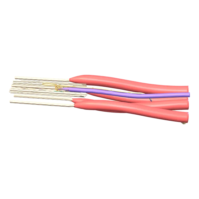
Palisade ending
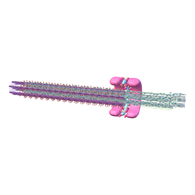
Sarcomere
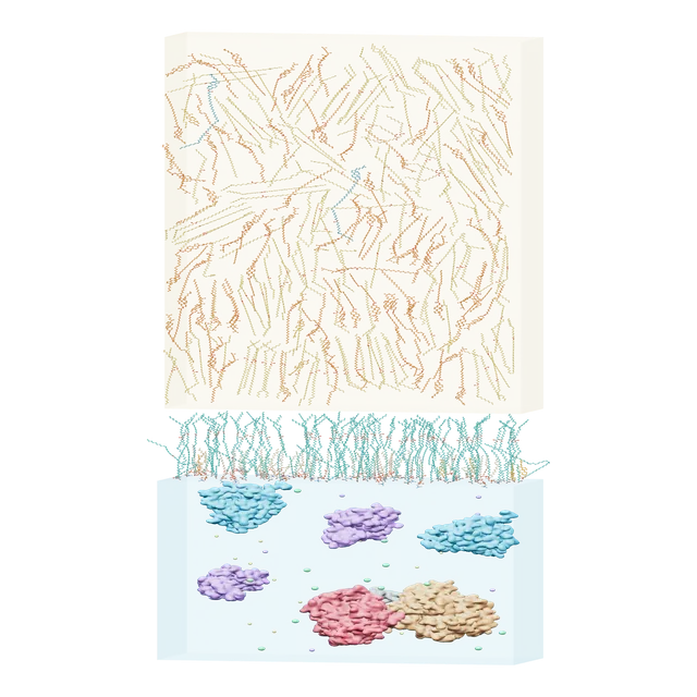
Tear lipid layer
Conjunctival surface glycocalyx
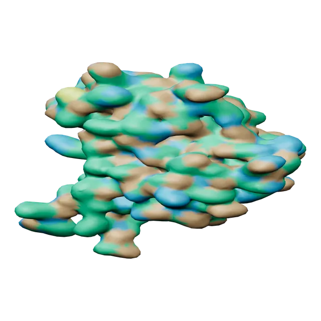
MUC16 SEA domain
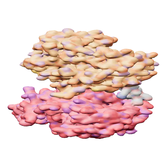
Lactoferrin
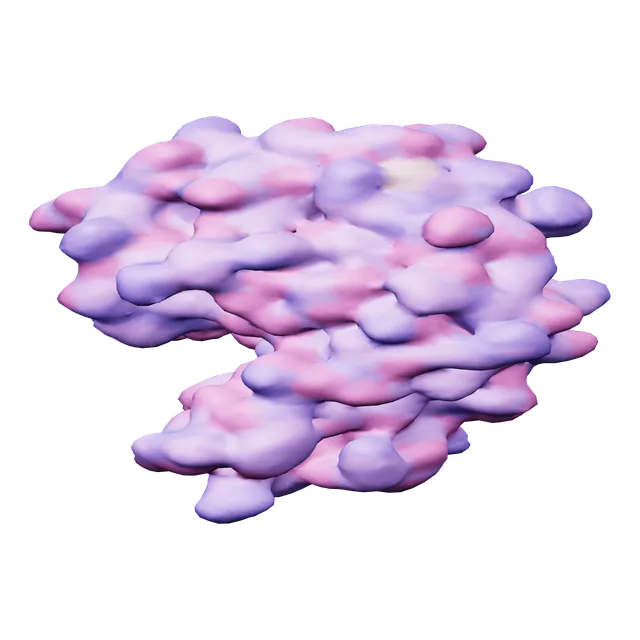
Lysozyme
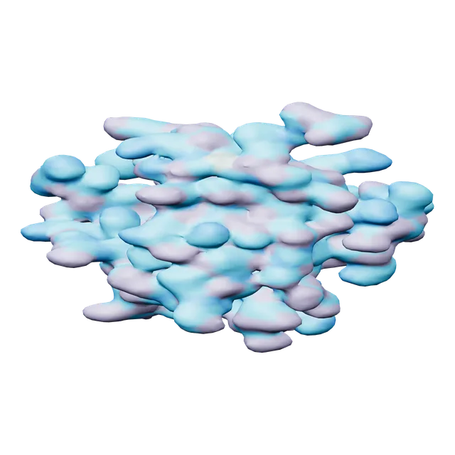
Tear lipocalin
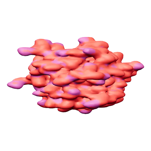
Galectin-3: carbohydrate recognition domain
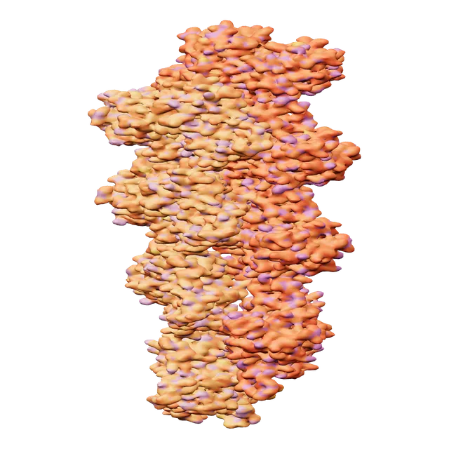
Actin filament
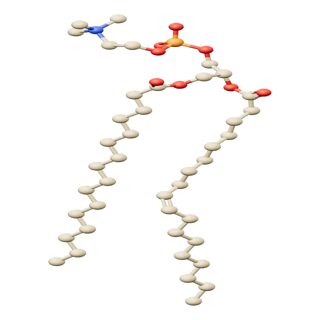
Phosphatidylcholine (POPC)
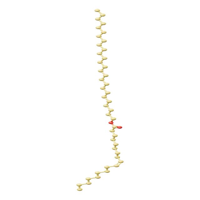
Wax ester
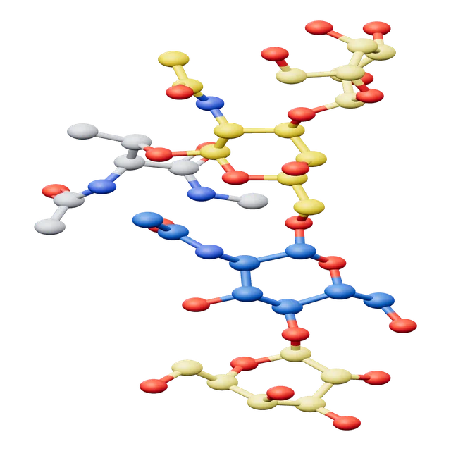
Branched Core 2 glycan
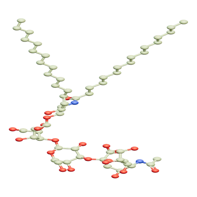
GM3 ganglioside
See the detail for yourself, and choose the level of display that fits your project.
Motion · 10 s previews
Watch the preview, then request the full video on WhatsApp.
BenefitShow your scientific mechanism in a clip made for conferences and social media.
IDRAK 3D · Patient education
The same atlas models, tailored to the clinic screen and simplified to show parents their child's eye, the problem and the doctor's plan.
BenefitClearer explanations help patients and families understand the condition and stick to treatment, and save the doctor's time.
Show how six muscles move each eye.
An illustrative simulation of vision before and after glasses helps parents see why they matter.
The child's face and the inside of both eyes on one screen, to explain strabismus to parents.
Walk through the surgical steps to discuss them with the patient and family.
With a single HDMI cable, families watch the explanation on a large screen.
Choose, pause and rotate a scene without leaving your seat.
Show content offline after activation, and connect to download updates.
Encrypted content, with a hidden watermark to protect its rights.
Updates arrive in the background when you're online. If one fails, the app rolls back to the previous version.
Educational content with no patient files or data.
For education and explanation only · Not a medical device · Does not diagnose · The decision is always the doctor'sAn app that helps patients and families understand.
Egyptian
Emirati
English
Clearer explanations help patients and families understand the condition and stick to treatment, and save the doctor's time.
Country partnerships
One partner per country, with a version in the local dialect and an annual licence for clinics. We provide the content, dialect and updates; you sell to clinics in your country.
Start your project
Tell us the part you need and how you'll use it, via WhatsApp or the form.